Review
. 2024 Jun;79(6):1470-1484.
doi: 10.1111/all.16044. Epub 2024 Feb 3.

The skin microbiome in pediatric atopic dermatitis and food allergy

Affiliations
Review

The skin microbiome in pediatric atopic dermatitis and food allergy

Elizabeth Huiwen Tham et al. Allergy. 2024 Jun.

Abstract

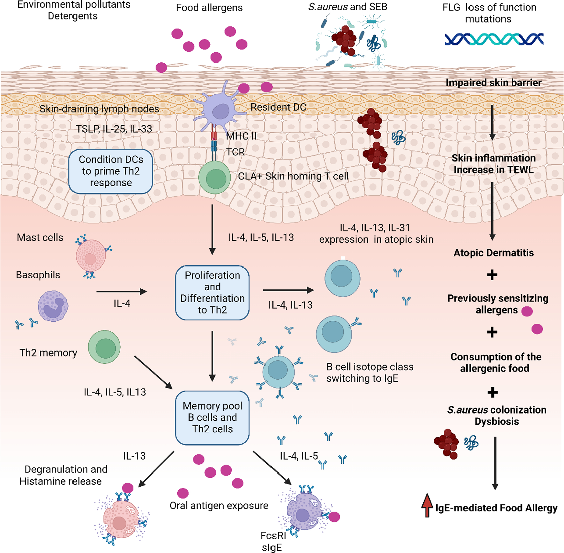
The skin microbiome is an extensive community of bacteria, fungi, mites, viruses and archaea colonizing the skin. Fluctuations in the composition of the skin microbiome have been observed in atopic dermatitis (AD) and food allergy (FA), particularly in early life, established disease, and associated with therapeutics. However, AD is a multifactorial disease characterized by skin barrier aberrations modulated by genetics, immunology, and environmental influences, thus the skin microbiome is not the sole feature of this disease. Future research should focus on mechanistic understanding of how early-life skin microbial shifts may influence AD and FA onset, to guide potential early intervention strategies or as microbial biomarkers to identify high-risk infants who may benefit from possible microbiome-based biotherapeutic strategies. Harnessing skin microbes as AD biotherapeutics is an emerging field, but more work is needed to investigate whether this approach can lead to sustained clinical responses.

Keywords: Staphylococcus aureus; atopic dermatitis; food allergy; microbiota; skin microbiome.


Conflict of interest statement

Conflict of Interest Statement

The authors declared no conflicts of interest

Figures

Figure 1:
Figure 1:. Interactions between Staphylococcus aureus, native skin commensals, and host cells in healthy or diseased skin.
On healthy skin (left), commensals like coagulase-negative Staphylococcus (CoNS) and the fungi Malassezia secrete a variety of compounds to inhibit the growth of the pathogen Staphylococcus aureus (S. aureus). CoNS secrete phenol soluble modulins (PSMs) and auto-inducing peptides, which inhibit S. aureus growth and virulence factor expression respectively. CoNS can stimulate host epidermal cells to produce anti-microbial peptides (AMPs) to further restrain S. aureus growth. Both CoNS and commensal Malassezia also secrete a variety of proteases, which disrupt S. aureus biofilm formation. These mechanisms contribute to T cell tolerance and likely optimize barrier function on healthy skin. It is however unclear if these proteases may also disrupt the host barrier to some extent. On inflamed skin (right), increased S. aureus colonization and biofilm formation can lead to heightened secretion of virulence factors such as PSMs, toxins and proteases that damage the stratum corneum. Host AMPs are present in lower amounts or are rendered less effective due to S. aureus activity and Th2 signalling. S. aureus can also suppress the growth or activity of skin commensals. Superantigens released by S. aureus can penetrate the epidermis and trigger dermal dendritic cells to drive T helper 2 (Th2) polarization and expansion. Excessive numbers of Th2 cells in turn, produce a variety of pro-inflammatory cytokines, which further exacerbate skin barrier dysfunction, IgE production by B-cells and mast cell degranulation. While the pre-flare skin (middle) is not inflamed, there might be decreases in the abundance of commensals which inhibit S. aureus, potentially promoting transition to a pathogenic state. These individuals have elevated Th2 responses and IgE compared to healthy skin, and are predisposed to severe itching in subsequent flares. (Created with BioRender.com)
Figure 2.
Figure 2.. Mechanisms of skin barrier dysfunction in atopic dermatitis resulting in food allergy.
Environmental pollutants, detergents, microbial dysbiosis, and genetics such as FLG loss of function mutations can lead to disruptions to the epithelial barrier. Skin barrier impairment leads to skin inflammation resulting in AD. Epicutaneous exposure to food allergens in the context of an impaired barrier may trigger food allergen sensitization through Th2-dependent pathways: specific resident dendritic cell (DC) subsets capture allergens in the skin and transport them to skin-draining lymph nodes where they are presented to the cell surface carbohydrate epitope cutaneous lymphocyte antigen (CLA+) skin homing T cells. IL‐4 and IL‐13 cytokines promote B‐cell isotype switching to specific IgE (sIgE), and upon differentiating into plasma cells, yield allergen‐specific IgE antibodies. The sIgE bind to high‐affinity FcεRI receptors on the surface of mast cells and basophils. During the sensitization process, a memory pool of allergen‐specific B cells and T helper 2 cells are produced. Upon subsequent oral antigen exposure (consumption of the allergenic food), cross-linking of sIgE on the FcεRI receptors trigger mast cell degranulation, release of histamine and other inflammatory mediators, culminating in the clinical allergic response. The presence of S. aureus enhances pro-inflammatory responses which heighten risk of FA development. S. aureus colonization, such as by methicillin-resistant S. aureus (MRSA), and exposure to staphylococcal enterotoxin B (SEB), has been shown to increase risk of FA independent of AD severity. S. aureus abundance was also found to be positively correlated with transepidermal water loss (TEWL) in patients with AD and FA. (Created with BioRender.com)

References

    1. Bylund S, Kobyletzki LB, Svalstedt M, et al. Prevalence and Incidence of Atopic Dermatitis: A Systematic Review. Acta Derm Venereol 2020; 100: adv00160. 2020/05/16. DOI: 10.2340/00015555-3510. - DOI - PMC - PubMed
    1. Langan SM, Irvine AD and Weidinger S. Atopic dermatitis. Lancet 2020; 396: 345–360. 2020/08/03. DOI: 10.1016/s0140-6736(20)31286-1. - DOI - PubMed
    1. Zheng TYJ, Hee Oh M, Zhu Z The atopic march: progression from atopic dermatits to allergic rhinitis and asthma. Allergy Asthma Immunol Res 2011; 3: 67–73. - PMC - PubMed
    1. Andersen YMFEA, Skov L, Thyssen JP Comorbidities of Atopic Dermatitis: Beyond Rhinitis and Asthma. Curr Derm Rep 2017; 6: 35–41. - PMC - PubMed
    1. Weidinger S, Beck LA, Bieber T, et al. Atopic dermatitis. Nat Rev Dis Primers 2018; 4: 1–1. 2018/06/23. DOI: 10.1038/s41572-018-0001-z. - DOI - PubMed

Publication types