

Diffractaic acid exerts anti-cancer effects on hepatocellular carcinoma HepG2 cells by inducing apoptosis and suppressing migration through targeting thioredoxin reductase 1
- PMID: 38308689
- PMCID: PMC11329542
- DOI: 10.1007/s00210-024-02980-5
Diffractaic acid exerts anti-cancer effects on hepatocellular carcinoma HepG2 cells by inducing apoptosis and suppressing migration through targeting thioredoxin reductase 1
Abstract
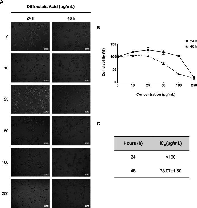
Hepatocellular carcinoma (HCC) represents one of the most common malignant tumors worldwide. Due to the limited number of available drugs and their side effects, the development of new chemotherapeutic strategies for HCC treatment has become increasingly important. This study is aimed at investigating whether diffractaic acid (DA), one of the secondary metabolites of lichen, exhibits a potential anticancer effect on HepG2 cells and whether its anticancer effect is mediated by inhibition of thioredoxin reductase 1 (TRXR1), which is a target of chemotherapeutic strategies due to overexpression in tumor cells including HCC. XTT assay results showed that DA exhibited strong cytotoxicity on HepG2 cells with an IC50 value of 78.07 µg/mL at 48 h. Flow cytometric analysis results revealed that DA displayed late apoptotic and necrotic effects on HepG2 cells. Consistent with these findings, real-time PCR results showed that DA did not alter the BAX/BCL2 ratio in HepG2 cells but upregulated the P53 gene. Moreover, the wound healing assay results revealed a strong anti-migratory effect of DA in HepG2 cells. Real-time PCR and Western blot analyses demonstrated that DA increased TRXR1 gene and protein expression levels, whereas enzyme activity studies disclosed that DA inhibited TRXR1. These findings suggest that DA has an anticancer effect on HepG2 cells by targeting the enzymatic inhibition of TRXR1. In conclusion, DA as a TRXR1 inhibitor can be considered an effective chemotherapeutic agent which may be a useful lead compound for the treatment of HCC.
Keywords: Cytotoxicity; Diffractaic acid; Expression; Hepatocellular carcinoma; Inhibition; Thioredoxin reductase 1.
© 2024. The Author(s).
Conflict of interest statement
The authors declare no competing interests.
Figures


Similar articles
-
Butaselen prevents hepatocarcinogenesis and progression through inhibiting thioredoxin reductase activity.Redox Biol. 2018 Apr;14:237-249. doi: 10.1016/j.redox.2017.09.014. Epub 2017 Sep 22. Redox Biol. 2018. PMID: 28965082 Free PMC article.
-
Diffractaic acid, a novel TrxR1 inhibitor, induces cytotoxicity, apoptosis, and antimigration in human breast cancer cells.Chem Biol Interact. 2022 Jul 1;361:109984. doi: 10.1016/j.cbi.2022.109984. Epub 2022 May 13. Chem Biol Interact. 2022. PMID: 35569514
-
Diffractaic acid exhibits thioredoxin reductase 1 inhibition in lung cancer A549 cells.J Appl Toxicol. 2023 Nov;43(11):1676-1685. doi: 10.1002/jat.4505. Epub 2023 Jun 17. J Appl Toxicol. 2023. PMID: 37329199
-
Antiproliferative, antimigratory, and apoptotic effects of diffractaic and vulpinic acids as thioredoxin reductase 1 inhibitors on cervical cancer.Naunyn Schmiedebergs Arch Pharmacol. 2024 Mar;397(3):1525-1535. doi: 10.1007/s00210-023-02698-w. Epub 2023 Sep 1. Naunyn Schmiedebergs Arch Pharmacol. 2024. PMID: 37658214
-
Da-Chai-Hu-Tang Formula inhibits the progression and metastasis in HepG2 cells through modulation of the PI3K/AKT/STAT3-induced cell cycle arrest and apoptosis.J Ethnopharmacol. 2024 Sep 15;331:118293. doi: 10.1016/j.jep.2024.118293. Epub 2024 May 3. J Ethnopharmacol. 2024. PMID: 38705430
Cited by
-
Inhibition of thioredoxin reductase 1 by evernic and vulpinic acids: a promising anticancer strategy on A549 cells.Naunyn Schmiedebergs Arch Pharmacol. 2025 Jun 11. doi: 10.1007/s00210-025-04363-w. Online ahead of print. Naunyn Schmiedebergs Arch Pharmacol. 2025. PMID: 40498096
-
In silico identification of PPARγ agonists from diffractaic acid analogs in prostate cancer: a comprehensive computational approach.3 Biotech. 2025 Jul;15(7):219. doi: 10.1007/s13205-025-04376-5. Epub 2025 Jun 20. 3 Biotech. 2025. PMID: 40546399
-
Lobaric Acid Exhibits Anticancer Potential by Modulating the Wnt/β-Catenin Signaling Pathway in MCF-7 Cells.Pharmacol Res Perspect. 2025 Aug;13(4):e70142. doi: 10.1002/prp2.70142. Pharmacol Res Perspect. 2025. PMID: 40576246 Free PMC article.
-
Development and synthesis of diffractaic acid analogs as potent inhibitors of colorectal cancer stem cell traits.Sci Rep. 2025 Feb 25;15(1):6695. doi: 10.1038/s41598-025-90552-9. Sci Rep. 2025. PMID: 40000756 Free PMC article.
References
-
- Branco V, Godinho-Santos A, Gonçalves J et al (2014) Mitochondrial thioredoxin reductase inhibition, selenium status, and Nrf-2 activation are determinant factors modulating the toxicity of mercury compounds. Free Radic Biol Med 73:95–105. 10.1016/j.freeradbiomed.2014.04.030 10.1016/j.freeradbiomed.2014.04.030 - DOI - PubMed
Publication types
MeSH terms
Substances
Grants and funding
LinkOut - more resources
Full Text Sources
Medical
Research Materials
Miscellaneous
