. 2024 Aug;131(8):985-997.
doi: 10.1016/j.ophtha.2024.01.035. Epub 2024 Feb 1.

A Retrospective Longitudinal Study of 460 Patients with ABCA4-Associated Retinal Disease

Affiliations

A Retrospective Longitudinal Study of 460 Patients with ABCA4-Associated Retinal Disease

Beau J Fenner et al. Ophthalmology. 2024 Aug.

Abstract

Purpose: To investigate the distribution of genotypes and natural history of ABCA4-associated retinal disease in a large cohort of patients seen at a single institution.

Design: Retrospective, single-institution cohort review.

Participants: Patients seen at the University of Iowa between November 1986 and August 2022 clinically suspected to have disease caused by sequence variations in ABCA4.

Methods: DNA samples from participants were subjected to a tiered testing strategy progressing from allele-specific screening to whole genome sequencing. Charts were reviewed, and clinical data were tabulated. The pathogenic severity of the most common alleles was estimated by studying groups of patients who shared 1 allele. Groups of patients with shared genotypes were reviewed for evidence of modifying factor effects.

Main outcome measures: Age at first uncorrectable vision loss, best-corrected visual acuity, and the area of the I2e isopter of the Goldmann visual field.

Results: A total of 460 patients from 390 families demonstrated convincing clinical features of ABCA4-associated retinal disease. Complete genotypes were identified in 399 patients, and partial genotypes were identified in 61. The median age at first vision loss was 16 years (range, 4-76 years). Two hundred sixty-five families (68%) harbored a unique genotype, and no more than 10 patients shared any single genotype. Review of the patients with shared genotypes revealed evidence of modifying factors that in several cases resulted in a > 15-year difference in age at first vision loss. Two hundred forty-one different alleles were identified among the members of this cohort, and 161 of these (67%) were found in only a single individual.

Conclusions: ABCA4-associated retinal disease ranges from a very severe photoreceptor disease with an onset before 5 years of age to a late-onset retinal pigment epithelium-based condition resembling pattern dystrophy. Modifying factors frequently impact the ABCA4 disease phenotype to a degree that is similar in magnitude to the detectable ABCA4 alleles themselves. It is likely that most patients in any cohort will harbor a unique genotype. The latter observations taken together suggest that patients' clinical findings in most cases will be more useful for predicting their clinical course than their genotype.

Financial disclosure(s): Proprietary or commercial disclosure may be found in the Footnotes and Disclosures at the end of this article.

Keywords: ABCA4; Cone–rod dystrophy; Retinitis pigmentosa; Severe juvenile-onset retinal dystrophy; Stargardt disease.


Conflict of interest statement

Conflicts of interest: there are no conflicting relationships for any of the authors.

Figures
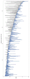
Figure 1 -
Figure 1 -. Proportion of each study participant’s symptomatic life for which ophthalmic records at the University of Iowa were available.
Subjects are ordered along the vertical axis by age at their baseline visit to the ophthalmology clinic at the University of Iowa. The years between their first uncorrectable vision loss and their baseline visit are shown as a gray bar, and the time between their baseline visit and their most recent visit is shown as a blue bar. To enhance legibility, blue bars for individuals are plotted with a minimum of one year in length even if patients were followed for a shorter interval.
Figure 3 -
Figure 3 -
Cumulative distribution of the age at which patients experienced their first uncorrectable vision loss.
Figure 4 -
Figure 4 -. Clinical Progression of Severe Juvenile Onset Retinal Dystrophy.
Fundus photographs (A-C), optical coherence tomograms (OCTs; D-F) and Goldmann visual fields (G-I) from the right eye of patient P312, at ages 8 years (best corrected visual acuity 5/200; A, D, G), 14 years (10/600; B), 15 years (20/1000; E, H,) and 20 years (20/800; C, F, I). At age 8, there is diffuse loss of outer retina leading to wrinkling of the internal limiting membrane, narrowing of the macular arterioles and loss of the I2e isopter. At ages 14-15, some bone-spicule-like pigmentation is seen, some gliotic thickening of the retina is evident on OCT and the I4e isopter is reduced to a small inferotemporal island. At age 20, there is extensive bone-spicule-like pigmentation, loss of the outer retinal laminations on OCT and an absolute scotoma 50 degrees in horizontal diameter. The absence of yellow flecks at all visits is indicative of the early and profound loss of photoreceptors. The OCT scale bars are 200 microns in both dimensions.
Figure 5 -
Figure 5 -. Clinical Variation of Bull’s Eye Maculopathy in Stargardt Disease.
Fundus photographs of nine individuals whose age at first vision loss was between 8 and 28 years: (A) left eye of patient P423, a 12 year old male with 20/100 acuity (Thr1526Met / IVS30+2028); (B) right eye of patient P151, a 47 year old woman with 20/125 acuity (Gly1961Glu / Cys2150Tyr); (C) right eye of patient P204, a 24 year old woman with 20/125 acuity (Gly863Ala/Asn1868Ile / Ala1598Asp); (D) right eye of patient P441, a 22 year old man with 20/20 acuity (Tyr245Stop / Not Identified); (E) right eye of patient P73, a 29 year old man with 20/80 acuity (Arg2107His / Gln1513Arg); (F) right eye of patient P412, a 26 year old woman with 20/250 acuity (Pro1380Leu / Phe418Ser); (G) left eye of patient P351, a 16 year old male with 10/125 acuity (IVS40+5 / Asp507Tyr); (H) right eye of patient P202, a 12 year old male with 20/50 acuity (Gly863Ala/Arg572Gln/Asn1868Ile / Trp821Arg); (I) right eye of patient P146, a 16 year old male with 20/160 acuity (Gly1961Glu / Asn1442Lys).
Figure 6 -
Figure 6 -. Clinical Progression of Autosomal Recessive Pattern Dystrophy.
Fundus photographs (A, B), optical coherence tomograms (OCTs; C, D) and Goldmann visual fields (E, F) from the right eye of patient P457, at ages 58 years (20/20; A, C, E), and 69 years (20/40; B, D, F). At age 58, coarse, reticular, yellow flecks are present in the macula that are visible subretinally on OCT. The visual field is full. At age 69, there is a zone of RPE and outer retinal atrophy nearly 5 mm in diameter with a small peninsula of foveal sparing. The I2e isopter has been almost completely lost but the I4e remains near normal. The OCT scale bars are 200 microns in both dimensions.
Figure 7 -
Figure 7 -. Age at first uncorrectable vision loss for individuals with one of the ten most common alleles in the cohort.
Blue vertical bars indicate the median age at first vision loss for each group. Each dot represents a single patient, and some patients appear in multiple rows.
Figure 8 -
Figure 8 -. Age at first uncorrectable vision loss for patients who share genotypes composed of the ten most common alleles.
Blue vertical bars indicate the median age at first vision loss for each group.
Figure 9 -
Figure 9 -. Clinically Discordant Individuals with Shared Genotype.
Fundus photos of two unrelated individuals who share the Pro1380Leu/IVS40+5 genotype. Panel A is a fundus photograph of the left eye of a 21 year old woman (patient P363; 20/125) who first experienced uncorrectable vision loss at age 15. She has nearly confluent flecks in the center of her macula and numerous more pisciform flecks extending superiorly beyond the arcades. Panel B is a fundus photograph of the right eye of a 61 year old woman (patient P361; 20/125) who first experienced uncorrectable vision loss at age 51. She has macula atrophy with peninsular foveal sparing and indistinct reticular flecks extending beyond the arcades.
Figure 10 -
Figure 10 -. Kaplan-Meier survival curves for visual function.
The gray line represents the survival curve for loss of the I2e isopter (less than or equal to the area of the optic disk). The blue line represents visual acuity worsening to a logMAR of ≥ 1. Vertical ticks along the lines represent censoring events. 95% confidence intervals are shown with shading.
Figure 11 -
Figure 11 -. Two Examples of Discordance in Visual Field and Visual Acuity Loss.
Fundus photographs (A, C), and Goldmann visual fields (B, D) from the right eyes of two individuals who met one Kaplan Meier severity criterion (Figure 9) more than 10 years before meeting the other; and, who each met the criteria in a different order. Patient P397 is a 29 year old woman with 20/125 acuity who met the logMAR >1 criterion at age 32 but still had a robust I2e isopter at that visit. Patient P101 is a 34 year old man with 20/30 acuity who met the criterion of a I2e isopter of less than 1 disk area on his first visit with us but did not meet the logMAR >1 criterion until eleven years later at age 45.

References

    1. Liew G, Michaelides M, Bunce C. A comparison of the causes of blindness certifications in England and Wales in working age adults (16-64 years), 1999-2000 with 2009-2010. BMJ Open 2014;4(2):e004015. - PMC - PubMed
    1. Heath Jeffery RC, Mukhtar SA, McAllister IL, et al. Inherited retinal diseases are the most common cause of blindness in the working-age population in Australia. Ophthalmic Genet 2021;42(4):431–9. - PMC - PubMed
    1. Stone EM, Andorf JL, Whitmore SS, et al. Clinically Focused Molecular Investigation of 1000 Consecutive Families with Inherited Retinal Disease. Ophthalmology 2017;124(9):1314–31. - PMC - PubMed
    1. Molday LL, Rabin AR, Molday RS. ABCR expression in foveal cone photoreceptors and its role in Stargardt macular dystrophy. Nat Genet 2000;25(3):257–8. - PubMed
    1. Lenis TL, Hu J, Ng SY, et al. Expression of ABCA4 in the retinal pigment epithelium and its implications for Stargardt macular degeneration. Proc Natl Acad Sci U S A 2018;115(47):E11120–E7. - PMC - PubMed

Substances