. 2024 Mar;300(3):105712.
doi: 10.1016/j.jbc.2024.105712. Epub 2024 Feb 1.

Rat hepatocytes secrete free oligosaccharides

Affiliations

Rat hepatocytes secrete free oligosaccharides

Chengcheng Huang et al. J Biol Chem. 2024 Mar.

Abstract

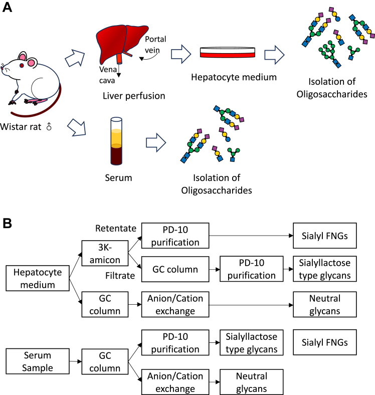
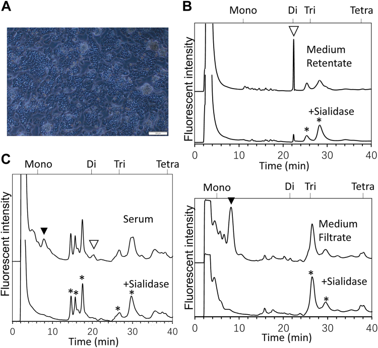
We recently established a method for the isolation of serum-free oligosaccharides, and characterized various features of their structures. However, the precise mechanism for how these glycans are formed still remains unclarified. To further investigate the mechanism responsible for these serum glycans, here, we utilized rat primary hepatocytes to examine whether they are able to secrete free glycans. Our findings indicated that a diverse array of free oligosaccharides such as sialyl/neutral free N-glycans (FNGs), as well as sialyl lactose/LacNAc-type glycans, were secreted into the culture medium by primary hepatocytes. The structural features of these free glycans in the medium were similar to those isolated from the sera of the same rat. Further evidence suggested that an oligosaccharyltransferase is involved in the release of the serum-free N-glycans. Our results indicate that the liver is indeed secreting various types of free glycans directly into the serum.

Keywords: free N-glycans; hepatocytes; milk oligosaccharides; oligosaccharyltransferase (OST); sialyl free oligosaccharides (glycans).


Conflict of interest statement

Conflict of interest The authors declare that they have no conflicts of interest with the contents of this article.

Figures

Figure 1
Figure 1
Flowchart for the isolation of primary hepatocytes and the detection of free glycans in the serum and the medium sample.A, scheme of this study. Male Wistar rats of 6 to 8 weeks old were used, and the primary hepatocytes were isolated as described in “Experimental procedures”. The culture medium for primary hepatocytes and serum were collected, and the free glycans were isolated from both the medium and the serum samples for analysis. B, flowchart for the isolation of free glycans. Top: method for the isolation of free glycans from the hepatocyte medium; bottom: method for the isolation of free glycans from serum (4).
Figure 2
Figure 2
Sialyl free glycans were detected in both the medium and the serum sample obtained from the same rat.A, representative image of the isolated primary hepatocytes (second day culture). The scale bar represents 200 μm. B, (top) DEAE anion exchange HPLC profile of the free sialyl FNGs (“Medium retentate” fraction) obtained from the second day culture medium of primary hepatocytes, and the sialidase-treated control; (bottom) DEAE anion exchange HPLC profile of the sialyl lactose/LacNAc-type free glycans (“Medium filtrate” fraction) obtained from the second day culture medium of primary hepatocytes, and the sialidase-treated control. C, DEAE anion exchange HPLC profile of the serum-free glycans obtained from the same rat. Open arrowhead indicates the sialidase-sensitive FNGs, and black arrowhead indicates the sialyl lactose/LacNAc-type free glycans. Asterisks marked in sialidase treated samples in B and C represent the peaks that are assumed to be nonspecific ones as they are resistant to sialidase. Moreover, no glycan-related signal was detected when the peaks between 12 to 18 min in C were collected and subjected for MS analysis. The neutral products are not detected in this HPLC system. DEAE, diethylaminoethyl cellulose; FNGs, free N-glycans.
Figure 3
Figure 3
Sialyl free glycans from serum and medium share same structures.A, dual gradient ODS HPLC profile of sialyl lactose/LacNAc-type free glycans collected form medium and serum samples. The sialidase treated control for the fractions was shown in Fig. S1. Peaks indicated by a and b represent the sialyl glycans whose structures could not be identified by combination of MS, enzyme reactions, as well as their elution positions in HPLC: predicted composition of each peak is as follows: a, NeuAc-Hex2; b, AcO-NeuAc(α2-3)-Hex2. Octothorpes represent the minor sialyl small glycans whose structures have not been determined due to its low amount. B, dual gradient ODS HPLC of disialyl FNGs collected from the medium and serum samples. L1-7, S1, and S2 marked in gray bars indicate the sialyl glycans detected in the medium and serum samples. Asterisks represent the fractions that are shown to be non-sialyl-glycan derived content after the fraction was collected for further examination by enzyme reaction. Octothorpes represent the fractions whose structures have not been identified, as its elution position on a dual gradient ODS HPLC does not match to standard samples available to us. The numbers above the chart are the elution positions of standard glucose oligomers with the number of glucose units (GU). Glycan structures and sugar compositions of L1-7, S1, and S2 were indicated in Table 1. FNGs, free N-glycans; ODS, octadecyl-silica.
Figure 4
Figure 4
Size fractionation HPLC profile of oligomannose-type FNGs from the serum and medium samples. HPLC profiles of the neutral fractions isolated from the serum (upper) and the medium (lower) samples and the jack bean α-mannosidase (JB Man’ase)-treated controls. Open arrowhead indicates the elution position of Man1GlcNAc1-PA, and black arrowhead indicates the elution position of Man1GlcNAc2-PA. Asterisks indicate the peaks that are most possibly non-glycan-derived, as they are resistant to various glycosidase digestions (glucoamylase, JB Man’ase, β-HexNAc’ase, as well as bovine Gal’ase). Peaks N1-9 marked in gray bars indicate the JB Man’ase-sensitive oligomannose-type glycans detected in the medium and serum samples. Sugar compositions indicated in Table 2 were estimated based on the elution positions of the samples on the size fractionation HPLC and the comparison with those of the authentic, previously characterized oligo/high mannose-type glycans (4, 36). The numbers above the chart are the elution positions of standard glucose oligomers with indicated glucose number as glucose unit (GU). FNG, free N-glycan.
Figure 5
Figure 5
OST is involved in the release of FNGs into the medium.A, DEAE anion exchange chromatography of the sialyl FNGs fractions from the medium of primary hepatocytes. To the medium the following compounds were separately added 1 day after the cells became attached to the dish: DMSO (vehicle control), 5 μM NGI-1(NGI-1), 0.5 mM castanospermine (CST), and 5 μM NGI-1/0.5 mM castanospermine (CST/NGI-1). The inhibitors were incubated overnight and the culture media were collected for free glycan analysis. Peaks marked in gray indicate the disialyl FNGs. B, size fractionation HPLC (upper) of the major sialyl FNGs in disialylated fractions after sialidase digestion and the calculation of the Gn1/Gn2 ratio (lower). The disialylated fractions (marked in gray in Fig. 5A) were collected, and treated with sialidase before the HPLC analysis. The Gn1+Gn2 glycans in the control was set to 1 and the relative amount in each fraction was calculated. C, size fractionation HPLC of the neutral fractions from control (Con) and NGI-1 treated (NGI-1) medium sample. The mannosidase-sensitive peaks that are decreased by NGI-1 treatment are indicated by open arrowheads. The mannosidase-sensitive peaks that are not changed after NGI-1 treatment are marked as black arrowheads. Asterisks indicate the peaks most possibly non-glycan derived, as they are resistant to glucoamylase, JB Man’ase, β-HexNAc’ase, as well as bovine Gal’ase. The samples were fractionated for small glycans (GU<5) and large glycans (GU>5), and further applied to the dual gradient ODS HPLC for the quantification of the change of the glycans after NGI-1 treatment in Fig. S5. D, calculation of the relative amount of mannose type FNGs after the NGI-1 treatment. The relative amount of Gn1 and Gn2 type high/oligomannose-type glycans was calculated as a total sum of Man1Gn1 and Man1Gn2 glycans, respectively, after JB Man’ase treatment as indicated by black arrowheads in Fig. S5, and the amount of Gn1 glycans in control sample (without inhibitors) was set to 1. Individual data points were presented, and error bars indicate SD from at least three independent experiments. Two-way ANOVA test was performed for Gn1 and Gn2 glycans to see the influence of inhibitor treatments. The p value was calculated by Turkey’s post hoc test, while N.S. represent not significant (i.e. p > 0.05). The numbers above the chart are the elution positions of standard glucose oligomers with indicated glucose number as glucose unit (GU). The use of monosaccharide symbols followed the Symbol Nomenclature for Glycans system (73), blue square, GlcNAc; green circle, Man; yellow circle, Gal. DEAE, diethylaminoethyl cellulose; DMSO, dimethyl sulfoxide; FNGs, free N-glycans; ODS, octadecyl-silica; OST, oligosaccharyltransferase.
Figure 6
Figure 6
Proposed model for the secretion of OST-generated Gn2-type FNGs from the hepatocytes into serum. The free N-glycans (FNGs) are released by the hydrolytic activity of OST, and the glycans are transported into the cytosol and processed by cytosolic ENGase and Man2C1, and then transported into lysosomes for further degradation in normal cells (red arrows). In hepatocytes, some FNGs escape from the translocation into the cytosol, pass through the vesicular transport and are further processed by the enzymes in the Golgi to convert high-mannose type glycans into complex-type ones. Sialyl FNGs thus formed are eventually secreted outside the cells (large pink arrows). The use of monosaccharide symbols followed the Symbol Nomenclature for Glycans system (73), purple diamond, NeuAc; blue square, GlcNAc; green circle, Man; yellow circle, Gal; blue circle, Glc. ENG, endo-β-N-acetylglucosaminidase; FNGs, free N-glycans; Man2C1, α-mannosidase 2C1; OST, oligosaccharyltransferase.

References

    1. Yoshida Y., Furukawa J.I., Naito S., Higashino K., Numata Y., Shinohara Y. Quantitative analysis of total serum glycome in human and mouse. Proteomics. 2016;16:2747–2758. - PubMed
    1. Pismenetskaya I.U., Butters T.D. Serum glycomarkers of endoplasmic reticulum and lysosomal-endosomal system stress in human healthy aging and diseases. Ukr Biochem. J. 2017;89:59–70. - PubMed
    1. Furukawa J.I., Hanamatsu H., Yokota I., Hirayama M., Ando T., Kobayashi H., et al. Comprehensive glycomic approach reveals novel low-molecular-weight blood group-specific glycans in serum and cerebrospinal fluid. J. Proteome Res. 2021;20:2812–2822. - PubMed
    1. Huang C., Seino J., Fujihira H., Sato K., Fujinawa R., Sumer-Bayraktar Z., et al. Occurrence of free N-glycans with a single GlcNAc at the reducing termini in animal sera. Glycobiology. 2022;32:314–332. - PubMed
    1. Hanzawa K., Tanaka-Okamoto M., Murakami H., Mukai M., Takahashi H., Omori T., et al. Investigation of acidic free-glycans in urine and their alteration in cancer. Glycobiology. 2021;31:391–409. - PMC - PubMed

Publication types

Substances

LinkOut - more resources