. 2024 Feb 3;56(1):6.
doi: 10.1007/s00726-023-03360-8.

D-Amino acids differentially trigger an inflammatory environment in vitro

Affiliations

D-Amino acids differentially trigger an inflammatory environment in vitro

Siew Hwei Yap et al. Amino Acids. .

Abstract

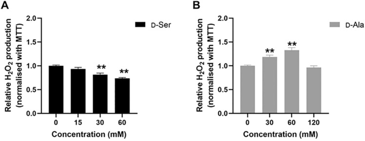
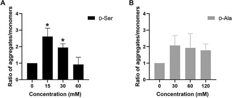
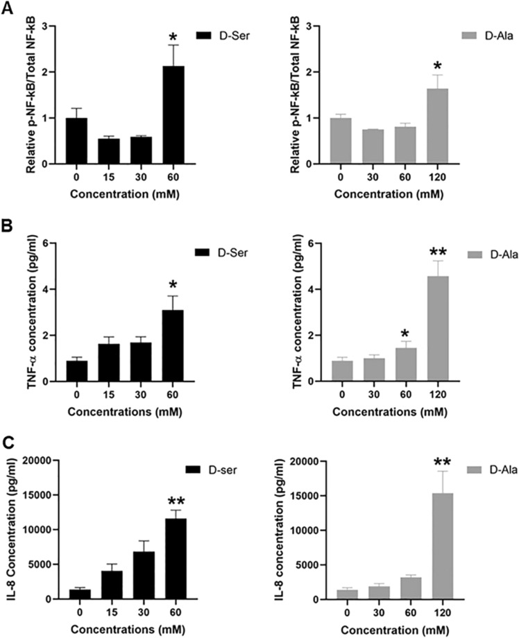
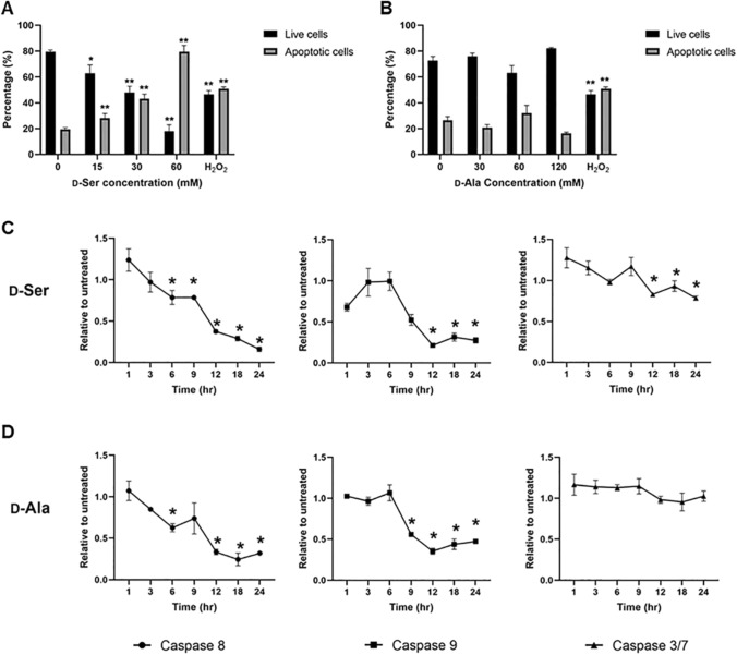
Studies in vivo have demonstrated that the accumulation of D-amino acids (D-AAs) is associated with age-related diseases and increased immune activation. However, the underlying mechanism(s) of these observations are not well defined. The metabolism of D-AAs by D-amino oxidase (DAO) produces hydrogen peroxide (H2O2), a reactive oxygen species involved in several physiological processes including immune response, cell differentiation, and proliferation. Excessive levels of H2O2 contribute to oxidative stress and eventual cell death, a characteristic of age-related pathology. Here, we explored the molecular mechanisms of D-serine (D-Ser) and D-alanine (D-Ala) in human liver cancer cells, HepG2, with a focus on the production of H2O2 the downstream secretion of pro-inflammatory cytokine and chemokine, and subsequent cell death. In HepG2 cells, we demonstrated that D-Ser decreased H2O2 production and induced concentration-dependent depolarization of mitochondrial membrane potential (MMP). This was associated with the upregulation of activated NF-кB, pro-inflammatory cytokine, TNF-α, and chemokine, IL-8 secretion, and subsequent apoptosis. Conversely, D-Ala-treated cells induced H2O2 production, and were also accompanied by the upregulation of activated NF-кB, TNF-α, and IL-8, but did not cause significant apoptosis. The present study confirms the role of both D-Ser and D-Ala in inducing inflammatory responses, but each via unique activation pathways. This response was associated with apoptotic cell death only with D-Ser. Further research is required to gain a better understanding of the mechanisms underlying D-AA-induced inflammation and its downstream consequences, especially in the context of aging given the wide detection of these entities in systemic circulation.

Keywords: D-Alanine; D-Amino acid oxidase; D-Serine; Inflammation; TNF-α.


Conflict of interest statement

The authors declare no competing interests.

Figures

Fig. 1
Fig. 1
d-Ser treatment reduced DAO mRNA expression and induced DAO protein expression, whereas treatment of d-Ala increased DAO mRNA expression and DAO protein expression in HepG2 cells. Gene expression levels of DAO were measured using RT-qPCR following 48 h of d-Ser (A) and d-Ala (B) treatment. Representative western blots and relative quantitative analysis of DAO in d-Ser-treated (C) and d-Ala-treated (D) cells. The data are expressed as the mean ± SEM (n = 3). Data were compared by the Student’s t-test. *p < 0.05 as compared to untreated cells
Fig. 2
Fig. 2
d-Ser treatment reduced hydrogen peroxide production but d-Ala treatment increased hydrogen peroxide production in HepG2 cells. The levels of hydrogen peroxide were measured by Amplex Red assay following 48 h of d-Ser (A) and d-Ala (B) treatment. The data are expressed as the mean ± SEM (n = 3). Data were compared by the Student’s t-test. **p < 0.001 as compared to untreated cells
Fig. 3
Fig. 3
Mitochondrial membrane potential decreased after 48 h of d-Ser treatment but not in d-Ala-treated cells. The membrane potential was measured using the red/green fluorescence ratio of the JC-1 dye in the mitochondria following 48 h of d-Ser (A) and d-Ala (B) treatment. Red shift of the dye means more aggregates are formed (higher membrane potential; hyperpolarization), whereas a lower red to green ratio means few aggregates are formed (lower membrane potential; depolarization). The ratios of aggregates/monomers were calculated relative to untreated samples. The data are expressed as the mean ± SEM (n = 3). Data were compared by the Student’s t test. *p < 0.05 as compared to untreated cells
Fig. 4
Fig. 4
Phosphorylated NF-κB protein expression, TNF-α, and IL-8 concentrations in cell supernatant were increased at the highest concentrations of d-Ser and d-Ala treatment. The levels of phosphorylated NF-κB protein (A) were determined by Western blotting after 48 h of d-Ser and d-Ala treatment. The levels of TNF-α (B) and IL-8 (C) were determined by Ella after 48 h of d-Ser and d-Ala treatment. The data are expressed as the mean ± SEM (n = 3). Data were compared by the Student’s t test. *p < 0.05, **p < 0.001 as compared to untreated cells
Fig. 5
Fig. 5
d-Ser induced a dose-dependent apoptosis in HepG2 cells but not d-Ala. Levels of caspase-8, 9, and 3/7 decreased in d-Ser treatment but only caspase-8 and 9 levels decreased in d-Ala treatment. Apoptosis was assessed using Annexin-V and propidium iodide (PI) kit after 48 h of d-Ser (A) and d-Ala (B) treatment using flow cytometry (see supplementary Fig. 3 for flow cytometry plots). Apoptotic cells were referred to as Annexin-V positive cells. Caspase-8, 9, and 3/7 activities were analyzed in HepG2 using Caspase-Glo kit over 24 h of d-Ser (C) and d-Ala (D) treatment. The data are expressed as the mean ± SEM (n = 3). Data were compared by the Student’s t-test. *p < 0.05, **p < 0.001 as compared to untreated cells
Fig. 6
Fig. 6
Schematic pathways for d-serine (d-Ser) and d-alanine (d-Ala) in HepG2 cells. In HepG2 cells, high concentrations of d-Ser activate general control nondepressible 2 (GCN2) instead of the DAO pathway, which induces the activation of NF-кB and the secretion of TNF-α, subsequently contributing to apoptosis. Although d-Ala did not cause apoptosis, the level of pro-inflammatory cytokine TNF-α increases through activation of NF-кB when HepG2 cells were treated with high concentrations of d-Ala

References

    1. Bardallo GR, Panisello-Roselló A, Sanchez-Nuno S, Alva N, Roselló-Catafau J, Carbonell T (2022) Nrf2 and oxidative stress in liver ischemia/reperfusion injury. FEBS J 289(18):5463–5479. 10.1111/febs.16336 - PubMed
    1. Brandish PE, Chiu C-S, Schneeweis J, Brandon NJ, Leech CL, Kornienko O, Scolnick EM, Strulovici B, Zheng W (2006) A cell-based ultra-high-throughput screening assay for identifying inhibitors of d-amino acid oxidase. J Biomol Screen 11(5):481–487. 10.1177/1087057106288181 - PubMed
    1. Buccitelli C, Selbach M (2020) mRNAs, proteins and the emerging principles of gene expression control. Nat Rev Genet 21(10):10. 10.1038/s41576-020-0258-4 - PubMed
    1. Chandel NS, Trzyna WC, McClintock DS, Schumacker PT (2000) Role of oxidants in NF-κB activation and TNF-α gene transcription induced by hypoxia and endotoxin. J Immunol 165(2):1013–1021. 10.4049/jimmunol.165.2.1013 - PubMed
    1. Chervyakov AV, Gulyaeva N, Zakharova M (2011) d-Amino acids in normal ageing and pathogenesis of neurodegenerative diseases. Neurochem J 8(2):113–129. 10.1134/S1819712411020036

LinkOut - more resources