. 2024 May;153(5):1355-1368.
doi: 10.1016/j.jaci.2024.01.017. Epub 2024 Feb 3.

Epithelial overexpression of IL-33 induces eosinophilic esophagitis dependent on IL-13

Affiliations

Epithelial overexpression of IL-33 induces eosinophilic esophagitis dependent on IL-13

Mia Y Masuda et al. J Allergy Clin Immunol. 2024 May.

Abstract

Background: Eosinophilic esophagitis (EoE) is an increasingly common inflammatory condition of the esophagus; however, the underlying immunologic mechanisms remain poorly understood. The epithelium-derived cytokine IL-33 is associated with type 2 immune responses and elevated in esophageal biopsy specimens from patients with EoE.

Objective: We hypothesized that overexpression of IL-33 by the esophageal epithelium would promote the immunopathology of EoE.

Methods: We evaluated the functional consequences of esophageal epithelial overexpression of a secreted and active form of IL-33 in a novel transgenic mouse, EoE33. EoE33 mice were analyzed for clinical and immunologic phenotypes. Esophageal contractility was assessed. Epithelial cytokine responses were analyzed in three-dimensional organoids. EoE33 phenotypes were further characterized in ST2-/-, eosinophil-deficient, and IL-13-/- mice. Finally, EoE33 mice were treated with dexamethasone.

Results: EoE33 mice displayed ST2-dependent, EoE-like pathology and failed to thrive. Esophageal tissue remodeling and inflammation included basal zone hyperplasia, eosinophilia, mast cells, and TH2 cells. Marked increases in levels of type 2 cytokines, including IL-13, and molecules associated with immune responses and tissue remodeling were observed. Esophageal organoids suggested reactive epithelial changes. Genetic deletion of IL-13 in EoE33 mice abrogated pathologic changes in vivo. EoE33 mice were responsive to steroids.

Conclusions: IL-33 overexpression by the esophageal epithelium generated immunopathology and clinical phenotypes resembling human EoE. IL-33 may play a pivotal role in the etiology of EoE by activating the IL-13 pathway. EoE33 mice are a robust experimental platform for mechanistic investigation and translational discovery.

Keywords: IL-33; eosinophil; eosinophilic esophagitis; transgene; type 2 inflammation.


Figures

Figure 1:
Figure 1:. A novel gene construct generated secreted and active IL-33.
(A) Secreted and active IL-33 (saIL-33) gene construct with a human CMV promoter (hCMV). (B) Immunocytochemical staining of IL-33 (red) on HEK-293 after transfection with either saIL-33 or empty vector. (C) Western blot of IL-33 in HEK-293 cell lysates after transfection with saIL-33 (lane 2) or empty vector (lane 3). Recombinant IL-33 (rIL-33) (5 ng, lane 1) was used as a positive control. B-actin was used as a loading control. (D) ELISA of eosinophil-derived IL-13 following in vitro culture with conditioned media from transfected (vector vs. saIL-33) HEK-293 cells. Data is shown as mean ± SEM. **p<0.01
Figure 2:
Figure 2:. Expression of functional saIL-33 by the mouse esophageal epithelium.
(A) The Epstein–Barr virus-derived promoter, ED-L2, was used to drive expression of saIL-33 from esophageal epithelium to generate “EoE33” mice. (B) Representative hematoxylin & eosin (H&E (top) and IL-33 immunohistochemistry staining (brown; bottom) of esophageal cross sections from wild-type (WT), EoE33, and EoE33 × ST2−/− mice. (C) IL-33 ELISA of whole tissue homogenates from EoE33 vs. WT mice. n = 4 mice per group. Data is shown as mean ± SEM. **** p<0.0001. Scale bar = 200 μm. Inset scale bar = 100 μm.
Figure 3:
Figure 3:. EoE33 mice exhibited features pathognomonic of eosinophilic esophagitis.
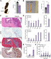
(A) Representative image and weights of eight-week-old male wild-type (WT) and EoE33 littermates. (B) Whole esophagi and esophageal diameter measurements from WT and EoE33 littermates. (C) Representative hematoxylin & eosin (H&E), eosinophil peroxidase (EPX (red)), Masson’s trichrome (MT), and Ki-67 (brown) staining of esophageal cross sections from WT and EoE33 mice. (D) H&E-stained esophageal biopsies from WT and EoE33 mice were assessed using the EoE Histologic Scoring System (EoEHSS). Esophageal cross sections were scored for both degree (grade) and extent (stage) of pathology. Mean EoEHSS component scores and composite scores are shown for EoE33 mice (WT scores = zero). EI, eosinophil infiltration, EA; eosinophil abscess, SL; eosinophil surface layering, DIS; dilated intracellular spaces; BZH; basal zone hyperplasia, and LPF; thickened lamina propria fibers. (E) H&E-stained esophageal biopsies from WT and EoE33 mice were assessed for peak eosinophils per high-power field (eos/hpf) in the epithelium and stroma. (F) Esophageal muscle tension in response to methacholine (MCh) in WT vs. EoE33 mice. n = 3 – 12 mice per group. Data is shown as mean ± SEM. **** p<0.0001, **p<0.01, *p<0.05. Scale bar = 100 μm. ND – not detected.
Figure 4:
Figure 4:. EoE33 mice exhibited esophageal type 2 inflammation and tissue remodeling.
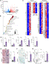
Bulk RNA-sequencing was performed on whole esophagi from eight-week-old male EoE33 and wild-type (WT) mice. (A) Volcano plot of differentially expressed genes (DEGs) between EoE33 and WT mice. Genes of interest are labeled. (B) Gene ontology (GO) analysis of up and downregulated DEG lists. Dot size represents the number of genes related to each biological process. (C) Heatmaps show normalized expression values for DEGs associated with immune and remodeling changes seen in EoE. The 20 largest absolute log2(fold change) values in each subset are bolded. (D) Type 2 cytokine levels in whole esophageal homogenates of WT vs. EoE33 mice. (E) Chloroacetate esterase (CAE) staining and mean CAE+ cells per esophageal cross section in WT vs. EoE33 mice. Arrows indicate CAE+ cells. (F) Immunohistochemistry staining for CD4 (brown) and CD4+ cells per esophageal cross section in WT vs. EoE33 mice. (G) Flow cytometry plot and quantification of esophageal Th2-type CD4+ T cells in WT vs. EoE33 mice. Frequencies shown as percentage of live, CD45+, CD3+, CD4+ population. n =−4 – 8 mice per group. Data shown as mean ± SEM. ***p<0.001, **p<0.01, *p<0.05. Scale bar = 100 μm.
Figure 5:
Figure 5:. EoE33 mice developed a food-specific humoral response to wheat.
Serially diluted serum from eight-week-old wild-type (WT) and EoE33 littermates was measured for antibodies against wheat, corn, and soy protein extracts (components of rodent chow) by ELISA. n = 4 mice per group. Data is shown as mean ± SEM. *p<0.05. O.D. – optical density.
Figure 6:
Figure 6:. EoE33 mice exhibited epithelial alterations in esophageal organoid cultures.
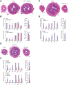
EoE33 and wild-type (WT) original esophageal epithelia (A) and organoids grown ex vivo (C-E) were subjected to morphological analyses. Representative hematoxylin & eosin (H&E) - stained and immunofluorescent (IF) images of esophageal epithelia (A) and organoids (C) with indicated genotypes. Esophageal epithelia were analyzed in longitudinal sections. Sox2 (red), Ki-67 (green), and DAPI (nuclei) are shown in IF. Bar graphs show Sox2 and Ki-67 labeling indices (LI) for original epithelia in (B) and organoids in (D). (E) WT esophageal organoids were treated with either IL-13 or IL-33 (10 ng/mL or 100 ng/mL) and representative H&E images are shown. Data is shown as mean ± SEM. Dashed line indicates basement membrane. Scale bar = 100 μm. ns – not significant.
Figure 7:
Figure 7:. EoE33 mice were responsive to steroid treatment and dependent on IL-13, whereas eosinophils were not required.
(A) Hematoxylin & eosin (H&E)-stained sections and (B) EoE Histologic Scoring System (EoEHSS) scores of wild-type (WT), EoE33, and eosinophil-deficient EoE33 (EoE33 × ΔdblGATA) mice. EI, eosinophil infiltration, EA; eosinophil abscess, ns; not significant, SL; eosinophil surface layering, DIS; dilated intracellular spaces; BZH; basal zone hyperplasia, and LPF; thickened lamina propria fibers. (C) H&E-stained sections and (D) EoEHSS scores of WT, EoE33, and IL-13-deficient EoE33 (EoE33 × IL-13−/−) mice. (E) H&E staining of representative esophageal cross sections from wild-type (WT) and EoE33 mice after dexamethasone treatment (dex) vs. saline (sal). (F) EoEHSS scores in dex-treated EoE33 mice vs. sal. n = 3 – 5 mice per group. Data is shown as mean ± SEM. Scale bar = 200 μm. *p< .05; **p< .01

References

    1. Rochman M, Azouz NP, Rothenberg ME. Epithelial origin of eosinophilic esophagitis. J Allergy Clin Immunol 2018; 142:10–23. - PMC - PubMed
    1. O’Shea KM, Aceves SS, Dellon ES, Gupta SK, Spergel JM, Furuta GT, et al. Pathophysiology of Eosinophilic Esophagitis. Gastroenterology 2018; 154:333–45. - PMC - PubMed
    1. Kottyan LC, Davis BP, Sherrill JD, Liu K, Rochman M, Kaufman K, et al. Genome-wide association analysis of eosinophilic esophagitis provides insight into the tissue specificity of this allergic disease. Nat Genet 2014; 46:895–900. - PMC - PubMed
    1. Simon D, Radonjic-Hosli S, Straumann A, Yousefi S, Simon HU. Active eosinophilic esophagitis is characterized by epithelial barrier defects and eosinophil extracellular trap formation. Allergy 2015; 70:443–52. - PubMed
    1. Judd LM, Heine RG, Menheniott TR, Buzzelli J, O’Brien-Simpson N, Pavlic D, et al. Elevated IL-33 expression is associated with pediatric eosinophilic esophagitis, and exogenous IL-33 promotes eosinophilic esophagitis development in mice. Am J Physiol Gastrointest Liver Physiol 2016; 310:G13–25. - PubMed

Publication types

MeSH terms