Review
. 2024 Feb 4;12(1):7.
doi: 10.1038/s41413-023-00304-6.

Spatial analysis of the osteoarthritis microenvironment: techniques, insights, and applications

Affiliations
Review

Spatial analysis of the osteoarthritis microenvironment: techniques, insights, and applications

Xiwei Fan et al. Bone Res. .

Abstract

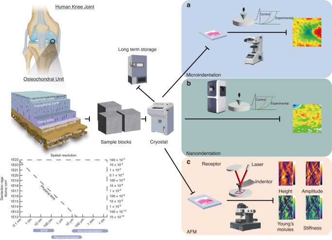
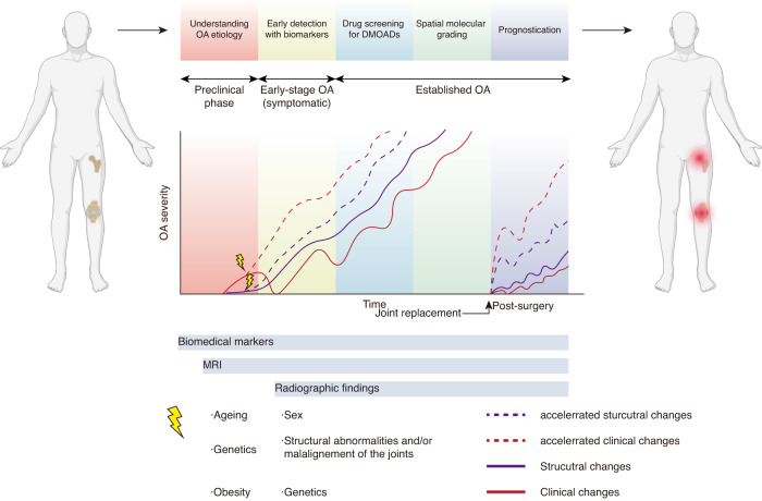
Osteoarthritis (OA) is a debilitating degenerative disease affecting multiple joint tissues, including cartilage, bone, synovium, and adipose tissues. OA presents diverse clinical phenotypes and distinct molecular endotypes, including inflammatory, metabolic, mechanical, genetic, and synovial variants. Consequently, innovative technologies are needed to support the development of effective diagnostic and precision therapeutic approaches. Traditional analysis of bulk OA tissue extracts has limitations due to technical constraints, causing challenges in the differentiation between various physiological and pathological phenotypes in joint tissues. This issue has led to standardization difficulties and hindered the success of clinical trials. Gaining insights into the spatial variations of the cellular and molecular structures in OA tissues, encompassing DNA, RNA, metabolites, and proteins, as well as their chemical properties, elemental composition, and mechanical attributes, can contribute to a more comprehensive understanding of the disease subtypes. Spatially resolved biology enables biologists to investigate cells within the context of their tissue microenvironment, providing a more holistic view of cellular function. Recent advances in innovative spatial biology techniques now allow intact tissue sections to be examined using various -omics lenses, such as genomics, transcriptomics, proteomics, and metabolomics, with spatial data. This fusion of approaches provides researchers with critical insights into the molecular composition and functions of the cells and tissues at precise spatial coordinates. Furthermore, advanced imaging techniques, including high-resolution microscopy, hyperspectral imaging, and mass spectrometry imaging, enable the visualization and analysis of the spatial distribution of biomolecules, cells, and tissues. Linking these molecular imaging outputs to conventional tissue histology can facilitate a more comprehensive characterization of disease phenotypes. This review summarizes the recent advancements in the molecular imaging modalities and methodologies for in-depth spatial analysis. It explores their applications, challenges, and potential opportunities in the field of OA. Additionally, this review provides a perspective on the potential research directions for these contemporary approaches that can meet the requirements of clinical diagnoses and the establishment of therapeutic targets for OA.


Conflict of interest statement

The authors declare no competing interests.

Figures

Fig. 1
Fig. 1
Magnitude and spatial resolution of the different instruments involved in deep spatial phenotyping. a Correlation map between the analyzed area and the spatial resolution of the state-of-the-art spatial phenotyping techniques used in the OA field. b Correlation map between the detection range and spatial resolution of the different instruments used for spatial phenotyping technologies. c Basic principle for deep spatial phenotyping in OA. This figure was created with Biorender.com
Fig. 2
Fig. 2
Pipeline of the different spatial phenotyping technologies in OA based on vibrational spectroscopic imaging. a NIR reflects C-H, N-H, and O–H stretching and bending overtones from 12 500 to 4 000 cm−1 with a resolution of 100 μm. It is fast, nondestructive, and labor-saving but has a wideband and overlap between different peaks in complex tissue, causing challenges in the differentiation of the characteristic peaks. MIR/FTIR reflects molecular vibrational and rotational energy level transitions (4 000–400 cm−1) with a resolution of 10 μm. It is fast, accurate, and nondestructive and reflects the information of most organic matter. However, it has a low signal on bone, causing challenges in the differentiation of the characteristic peaks and complexes for the determination of the exact mass. b Based on the principle of Raman scattering, Raman spectrometry imaging reflects the vibrational information between molecules with a resolution of 1 μm–250 nm. It is fast, accurate, and nondestructive, reflecting the information of most organic matter. However, the optical system and fluorescence interference can alter the Raman scattering region. c Nano-FTIR combines FTIR with scattering-type scanning near-field optical microscopy (s-SNOM). Based on AFM, an external light source illuminates a sharp tip, and the tip-scattered light (usually backscattered) is measured as a function of tip position. Its spatial resolution can reach 20 nm. The advantages and disadvantages are similar to FTIR. Advanced techniques enable the investigation of the microenvironment of cells and tissues at a subcellular level. NIR: Near infrared, MWIR: Middle wave infrared, FIR: Far infrared. This figure was created with BioRender.com
Fig. 3
Fig. 3
Pipelines of the different multiomics spatial phenotyping technologies used in the OA field. a Spatial transcriptomics is a cutting-edge technology that merges oligonucleotides with slides and combines them with tissue samples for detection. This method enables the detection of RNA signals with high accuracy, sensitivity, and a resolution of 200 nanometers. However, this technology is relatively costly and has potentially compatibility challenges when applied to bone and cartilage specimens. b Spatial proteomics uses certain protein enzymes to dissect the protein sequence into peptide segments. The sample is then ionized using different techniques, including matrix-assisted laser desorption/ionization (MALDI) and desorption electrospray ionization (DESI); finally, the laser is used to extract the mass for mapping with a resolution up to 20 μm. It spatially detects peptide signals, with mid to high sensitivity. However, the data is difficult to interpret. Specific peptide signals point to various proteins, and they need further validation. c Mass spectrometry imaging (MSI)-guided multiomics combines MSI and laser microdissection microscopy for separation, cross-validation and analysis. It can reach high accuracy and sensitivity but requires an optimized section-transferring system. d Digital spatial profiler (DSP) employs barcoded fluorescent markers for RNA and protein detection. It is easy to use with multiple genes and protein detection capability available, although it shows only partial information in the gene clusters without mapping. e Functional mass spectrometry imaging (fMSI) uses specific substrates for digesting the enzyme. Then, MSI instruments can extract ionized product signals for mapping with a resolution of up to 20 μm, confirming high sensitivity and accuracy when spatially detecting substrates and products. However, researchers need to first validate the existence of the enzyme. This figure was created with BioRender.com
Fig. 4
Fig. 4
Pipelines of the different spatial phenotyping technologies used in elemental phenotyping imaging. a Laser ablation–inductively coupled plasma‒mass spectrometry (LA-ICP‒MS) imaging uses a laser to ablate the tissue, which is then delivered with a carrier gas to the inductively coupled plasma (ICP). The ICP atomizes the sample, and the mass spectrometer analyzer analyses the generated ions. ICP‒MS can analyze sizes as low as 1 μm and has a mass-specific ability to simultaneously monitor several elements. b Synchrotron-micro X-ray fluorescence (SR-μXRF) imaging uses synchrotron radiation as an excitation source. An inner shell electron of the atom is struck and ejected by the X-ray. Fluorescence radiation is emitted and measured once an electron from a further outer shell fills the vacant shell site with a spatial resolution of up to 1 μm. This technique enables quantitative measurements that spatially detect elements. c Time-of-flight secondary ion mass spectrometry (TOF-SIMS) imaging dislodges chemical species on a material’s surface using a focused, pulsed ion beam. The time of fight of the produced dissociated ions for elemental and some molecular components with spatial resolution as low as 50-100 nm are detected. The spatial resolution depends upon the focused ion beam used, which is contingent on the sample type being analyzed. It has the advantages of a wide detection range, high accuracy and sensitivity. The use of this technique for bone analysis can be challenging due to charge accumulation during analyses. However, new-generation instruments are more effectively mitigating charge buildup. The results are only semiquantitative, and companion XPS analysis can often improve the associated quantitation. This figure was created with BioRender.com
Fig. 5
Fig. 5
Pipelines of the different spatial phenotyping technologies used in elemental phenotyping imaging. a Microindentation enables basic elemental mapping using a microindenter. It is easy to use but has relatively low sensitivity and spatial resolution. b Nanoindentation enables an improved indentation function with a smaller gap between indentation points and can reach a spatial resolution of 100 μm. However, it has a lower imaging area compared with microindentation. c Atomic force microscopy (AFM) uses a cantilever as a nanoindenter with a spatial resolution of up to 1–5 nm. It enables mechanical testing at the molecular level. However, it requires a steep learning curve with a narrow window for imaging. This figure was created with BioRender.com
Fig. 6
Fig. 6
Landscape of clinical application in all stages of OA with deep spatial phenotyping techniques. Deep spatial phenotyping techniques can be used for understanding OA etiology, early detection with integrated biomarkers, drug screening for disease-modifying drugs for OA (DMOADs), spatial molecular grading, and prognostication of OA. This figure was created with BioRender.com

References

    1. Hawker GA. Osteoarthritis is a serious disease. Clin. Exp. Rheumatol. 2019;37(Suppl 120):3–6. - PubMed
    1. Leifer VP, Katz JN, Losina E. The burden of OA-health services and economics. Osteoarthr. Cartil. 2022;30:10–16. doi: 10.1016/j.joca.2021.05.007. - DOI - PMC - PubMed
    1. Kraus VB, Blanco FJ, Englund M, Karsdal MA, Lohmander LS. Call for standardized definitions of osteoarthritis and risk stratification for clinical trials and clinical use. Osteoarthr. Cartil. 2015;23:1233–1241. doi: 10.1016/j.joca.2015.03.036. - DOI - PMC - PubMed
    1. Felson DT, et al. The prevalence of knee osteoarthritis in the elderly. The Framingham Osteoarthritis Study. Arthritis Rheum. 1987;30:914–918. doi: 10.1002/art.1780300811. - DOI - PubMed
    1. Jordan JM, et al. Prevalence of knee symptoms and radiographic and symptomatic knee osteoarthritis in African Americans and Caucasians: the Johnston County Osteoarthritis Project. J. Rheumatol. 2007;34:172–180. - PubMed

Publication types