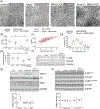
PMP2 regulates myelin thickening and ATP production during remyelination
- PMID: 38311982
- PMCID: PMC11027087
- DOI: 10.1002/glia.24508
PMP2 regulates myelin thickening and ATP production during remyelination
Abstract
It is well established that axonal Neuregulin 1 type 3 (NRG1t3) regulates developmental myelin formation as well as EGR2-dependent gene activation and lipid synthesis. However, in peripheral neuropathy disease context, elevated axonal NRG1t3 improves remyelination and myelin sheath thickness without increasing Egr2 expression or activity, and without affecting the transcriptional activity of canonical myelination genes. Surprisingly, Pmp2, encoding for a myelin fatty acid binding protein, is the only gene whose expression increases in Schwann cells following overexpression of axonal NRG1t3. Here, we demonstrate PMP2 expression is directly regulated by NRG1t3 active form, following proteolytic cleavage. Then, using a transgenic mouse model overexpressing axonal NRG1t3 (NRG1t3OE) and knocked out for PMP2, we demonstrate that PMP2 is required for NRG1t3-mediated remyelination. We demonstrate that the sustained expression of Pmp2 in NRG1t3OE mice enhances the fatty acid uptake in sciatic nerve fibers and the mitochondrial ATP production in Schwann cells. In sum, our findings demonstrate that PMP2 is a direct downstream mediator of NRG1t3 and that the modulation of PMP2 downstream NRG1t3 activation has distinct effects on Schwann cell function during developmental myelination and remyelination.
Keywords: FABP8; NRG1t3; PMP2; Schwann cell; myelin.
© 2024 The Authors. GLIA published by Wiley Periodicals LLC.
Figures


Similar articles
-
Axonal neuregulin 1 is a rate limiting but not essential factor for nerve remyelination.Brain. 2013 Jul;136(Pt 7):2279-97. doi: 10.1093/brain/awt148. Brain. 2013. PMID: 23801741 Free PMC article.
-
Akt Regulates Axon Wrapping and Myelin Sheath Thickness in the PNS.J Neurosci. 2016 Apr 20;36(16):4506-21. doi: 10.1523/JNEUROSCI.3521-15.2016. J Neurosci. 2016. PMID: 27098694 Free PMC article.
-
Neural Progenitor-Like Cells Induced from Human Gingiva-Derived Mesenchymal Stem Cells Regulate Myelination of Schwann Cells in Rat Sciatic Nerve Regeneration.Stem Cells Transl Med. 2017 Feb;6(2):458-470. doi: 10.5966/sctm.2016-0177. Epub 2016 Sep 7. Stem Cells Transl Med. 2017. PMID: 28191764 Free PMC article.
-
Transcription Factors and Coregulators in Schwann Cell Differentiation, Myelination, and Remyelination: Implications for Peripheral Neuropathy.J Neurosci Res. 2025 Jun;103(6):e70053. doi: 10.1002/jnr.70053. J Neurosci Res. 2025. PMID: 40452391 Review.
-
Neuregulin/ErbB Signaling in Developmental Myelin Formation and Nerve Repair.Curr Top Dev Biol. 2016;116:45-64. doi: 10.1016/bs.ctdb.2015.11.009. Epub 2016 Feb 1. Curr Top Dev Biol. 2016. PMID: 26970613 Review.
Cited by
-
Pmp2+ Schwann Cells Maintain the Survival of Large-Caliber Motor Axons.J Neurosci. 2025 Mar 26;45(13):e1362242025. doi: 10.1523/JNEUROSCI.1362-24.2025. J Neurosci. 2025. PMID: 39880678
-
Proteomic Analysis of Chronic Binge Alcohol-Induced Hippocampal and Anterior Cingulate Cortex Neuroadaptations in Simian Immunodeficiency Virus (SIV)-Infected Female Rhesus Macaques.J Neuroimmune Pharmacol. 2025 Feb 10;20(1):16. doi: 10.1007/s11481-025-10179-5. J Neuroimmune Pharmacol. 2025. PMID: 39930298 Free PMC article.
-
High-Fat Diet Disrupt Nerve Function by Targeting Schwann Cells.J Peripher Nerv Syst. 2025 Jun;30(2):e70036. doi: 10.1111/jns.70036. J Peripher Nerv Syst. 2025. PMID: 40522084
-
Peripheral myelin protein 2 is underexpressed in early-onset colorectal cancer and inhibits metastasis.Front Mol Biosci. 2025 Jun 5;12:1610003. doi: 10.3389/fmolb.2025.1610003. eCollection 2025. Front Mol Biosci. 2025. PMID: 40538623 Free PMC article.
-
Quantitative proteomics unveils known and previously unrecognized alterations in neuropathic nerves.J Neurochem. 2024 Sep;168(9):3154-3170. doi: 10.1111/jnc.16189. Epub 2024 Jul 29. J Neurochem. 2024. PMID: 39072727 Free PMC article.
References
-
- Acheta J, Hong J, Jeanette H, Brar S, Yalamanchili A, Feltri ML, Manzini MC, Belin S, & Poitelon Y. (2022). Cc2d1b contributes to the regulation of developmental myelination in the central nervous system. Frontiers in Molecular Neuroscience, 15, 881571. 10.3389/fnmol.2022.881571 - DOI - PMC - PubMed
Publication types
MeSH terms
Substances
Grants and funding
LinkOut - more resources
Full Text Sources
Molecular Biology Databases

