

Primary Pulmonary Synovial Sarcoma with Hemothorax: a Case Report
- PMID: 38313103
- PMCID: PMC10834050
- DOI: 10.5455/medarh.2023.77.496-499
Primary Pulmonary Synovial Sarcoma with Hemothorax: a Case Report
Abstract
Background: Synovial sarcoma is a rare and aggressive soft tissue malignancy most commonly arises from periarticular tissue of the extremities. Although several cases in the literature have reported different origins, primary pulmonary synovial sarcoma (PPSS) is an exceedingly rare and underrecognized entity, accounting for 0.5% of all lung malignancies. Clinical presentation includes chest pain, dyspnea, cough, and hemoptysis. The finding of hemothorax is a rare presentation and was barely reported in the literature. Due to its rarity and aggressive nature, the optimal treatment is unclear, while the mainstay remains surgical resection with chemo- and/or radiation therapy.
Objective: To report a case of hemorrhagic effusion subsequently diagnosed with primary pulmonary synovial sarcoma with the main objective of enriching the literature regarding this rare malignancy.
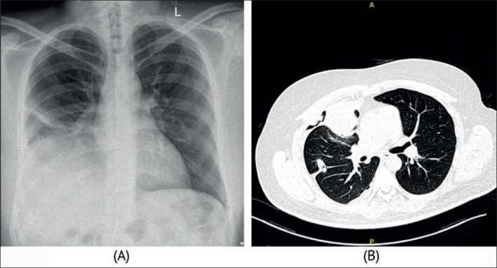
Case report: A 52-year-old male smoker with a background of coronary artery disease, hypertension, and diabetes mellitus was referred to our hospital. The patient presented with a history of chest pain, dyspnea, and massive right-sided pleural effusion. Laboratory investigations were unremarkable except for anemia. Chest x-ray showed a complete opacity on the right lower zone with right-sided pleural effusion. Thoracentesis was done and revealed hemorrhagic exudative effusion. Computed tomography (CT) scan showed a right heterogeneous lung mass compressing the medial segment of the middle lobe. Subsequently, the patient underwent bronchoscopy, which showed compression and edema on the right middle lobe bronchus with traces of blood coming from the right lower lobe. The patient underwent a right posterolateral thoracotomy, a fungating mass eroding the medial segment of the middle lobe was resected that was diagnosed as high-grade primary pulmonary synovial sarcoma. Radiotherapy was instituted. The patient died after two years due to recurrence.
Conclusion: PPSS is an aggressive disease with poor prognostic outcomes, and Its presentation is almost similar to other lung malignancies. Meanwhile, there is no definitive management guideline, and most management depends on surgical resection if feasible with adjuvant chemo-radiation therapy.
Keywords: Primary pulmonary synovial sarcoma; care report; hemothorax; lung cancer; lung sarcoma.
© 2023 Abdullah Abdulaziz AlQatari, Ayesha Ahmed, Fatima AlHije, Mohammed Sabry, Hatem Elbawab.
Conflict of interest statement
There were no conflicts of interest.
Figures


References
-
- Suster S, Moran C. Second. Philadelphia, PA: Elsevier; 2017. DIAGNOSTIC PATHOLOGY: THORACIC.
Publication types
MeSH terms
LinkOut - more resources
Full Text Sources
Medical