. 2023 Oct;16(10):1488-1498.
doi: 10.25122/jml-2023-0064.

Targeting VEGF using Bevacizumab attenuates sepsis-induced liver injury in a mouse model of cecal ligation and puncture

Affiliations

Targeting VEGF using Bevacizumab attenuates sepsis-induced liver injury in a mouse model of cecal ligation and puncture

Aula Zaini et al. J Med Life. 2023 Oct.

Abstract

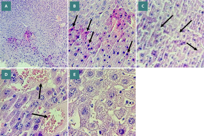
Sepsis, a life-threatening condition resulting from an uncontrolled host response to infection, often leads to severe liver damage and remains a significant cause of mortality in critically ill patients despite advances in antibiotic therapy and resuscitation. Bevacizumab, a neutralizing antibody targeting vascular endothelial growth factor (VEGF), is approved for treating certain cancers. However, its potential impact on sepsis-related liver injury is not well understood. This study aimed to explore the potential hepatoprotective effect of Bevacizumab on sepsis-induced liver injury. Twenty-four mice were divided into four groups: a sham group subjected to a midline incision only, a cecal ligation and puncture induction (CLP) group, a vehicle-treated group that received a vehicle one hour before CLP induction, and a Bevacizumab-treated group that received Bevacizumab one hour before CLP induction. Blood samples were collected, and angiopoietin-2 (ANGPT2), alanine transaminase (ALT), and aspartate transaminase (AST) serum levels were measured. Liver tissue homogenates were analyzed for IL-6, TNFα, intracellular adhesion molecule (ICAM-1), macrophage inhibitory factor (MIF), vascular endothelial growth factor (VEGF), F2-isoprostane, and caspase-11 levels. A histological examination was performed to assess the extent of liver damage. Mice exposed to CLP had high levels of the biomarkers mentioned above with a high degree of liver injury compared to the sham group. In contrast, treatment with Bevacizumab notably reduced these markers and mitigated liver damage. In conclusion, Bevacizumab may be a protective agent against sepsis-induced liver injury.

Keywords: Bevacizumab; VEGF; mouse model; sepsis-induced liver injury.


Conflict of interest statement

The authors declare no conflict of interest.

Figures

Figure 1
Figure 1
Effect of Bevacizumab on AST and ALT levels following CLP-induced liver injury Serum levels of AST and ALT across the four groups were measured in U/L. Data are presented as mean±SEM, n=6. One-way ANOVA and Bonferroni test.***p≤0.001, ****p≤0.0001
Figure 2
Figure 2
Effect of Bevacizumab on TNF-α and IL-6 levels following CLP Levels of TNF-α and IL-6 in the tissues of the liver across the four groups were measured in ng/L. Results are presented as mean±SEM, n=6. One-way ANOVA and Bonferroni test, **p≤0.01.
Figure 3
Figure 3
Effect of Bevacizumab on F2-isoprostane levels following CLP The levels of F2-isoprostane in the liver across the four groups were measured in ng/L. Results are presented as mean ±SEM, n=6. One-way ANOVA and Bonferroni test, ***p≤0.001.
Figure 4
Figure 4
Effect of Bevacizumab on caspase-11 levels following CLP The levels of caspase-11 in the liver across the four groups were measured in ng/L. The results are presented as mean±SEM, n=6. One-way ANOVA and Bonferroni test, **p≤0.01.
Figure 5
Figure 5
Effect of Bevacizumab on MIF levels following CLP The levels of MIF in the tissues of the liver across the four groups were measured in ng/L. The results are expressed as mean±SEM, n=6. One-way ANOVA and Bonferroni test, ****p≤0.0001.
Figure 6
Figure 6
Effect of Bevacizumab on ANGPT2 levels following CLP Serum levels of ANGPT2 across the four groups were measured in ng/L. Data are presented as mean±SEM, n=6. One-way ANOVA and Bonferroni test.***p≤0.001.
Figure 7
Figure 7
Effect of Bevacizumab on ICAM-1 levels following CLP ICAM-1 levels in the tissues of the liver across the four study groups were measured in ng/L. The results are presented as mean ±SEM, n=6. One-way ANOVA and Bonferroni test, ****p≤0.0001.
Figure 8
Figure 8
Effect of Bevacizumab on VEGF levels following CLP VEGF levels in liver tissues across the four study groups were measured in ng/L. The results are presented as mean±SEM, n=6. One-way ANOVA and Bonferroni test, ****p≤0.0001.
Figure 9
Figure 9
Histological liver damage scores Mean histological scores representing liver damage across four experimental groups. Data presented as mean±SEM, n=6. Kruskal-Wallis test ***p≤0.001.
Figure 10
Figure 10
Liver histology in sham group. A: Normal liver histology in the sham group (X 100 magnification). B: Central vein (red arrow) and normal hepatocytes (black arrow) in the sham group (X 400 magnification).
Figure 11
Figure 11
Liver sections showing the morphological changes in the CLP group. A: Severe liver damage in liver sections (score 3) (X100 magnification). B: Cytoplasmic eosinophilia, apoptotic cells with chromatin condensation, pyknotic nuclei (black arrows) (X400 magnification). C: Necrotic cells with nuclear fading (black arrows) (X400 magnification). D: Vascular congestion with erythrocyte stasis (black arrows) (X400 magnification). E: Cytoplasmic vacuoles (X400 magnification).
Figure 12
Figure 12
Liver sections showing the impact of Bevacizumab treatment on liver tissues following CLP. A: Mild liver damage in liver sections (score 1) (X100 magnification). B: Vascular congestion with erythrocyte stasis (red arrows) (X400 magnification). C: Vascular congestion with erythrocyte stasis (black arrows), cytoplasmic vacuoles, and steatosis (red arrows) (X400 magnification).

Similar articles

Cited by

References

    1. Zhu W, Bao R, Fan X, Tao T, et al. PD-L1 Blockade Attenuated Sepsis-Induced Liver Injury in a Mouse Cecal Ligation and Puncture Model. Mediators of Inflammation. Hindawi. 2013:e361501. doi: 10.1155/2013/361501. - DOI - PMC - PubMed
    1. Jeong SJ, Han SH, Kim CO, Choi JY, Kim JM. Anti-vascular endothelial growth factor antibody attenuates inflammation and decreases mortality in an experimental model of severe sepsis. Crit Care. 2013;17(3):R97. doi: 10.1186/cc12742. - DOI - PMC - PubMed
    1. Bauer M, Gerlach H, Vogelmann T, Preissing F, et al. Mortality in sepsis and septic shock in Europe, North America and Australia between 2009 and 2019-results from a systematic review and meta-analysis. Crit Care. 2020;24:239. doi: 10.1186/s13054-020-02950-2. - DOI - PMC - PubMed
    1. Wang X, Bove AM, Simone G, Ma B. Molecular Bases of VEGFR-2-Mediated Physiological Function and Pathological Role. Front Cell Dev Biol. 2020;8:599281. doi: 10.3389/fcell.2020.599281. - DOI - PMC - PubMed
    1. Rauniyar K, Jha SK, Jeltsch M. Biology of Vascular Endothelial Growth Factor C in the Morphogenesis of Lymphatic Vessels. Front Bioeng Biotechnol. 2018;6 doi: 10.3389/fbioe.2018.00007. - DOI - PMC - PubMed

MeSH terms

Substances