[Preprint]. 2024 Jan 18:rs.3.rs-3838282.
doi: 10.21203/rs.3.rs-3838282/v1.

Exposure to Gulf war illness-related chemicals exacerbates alcohol- induced liver damage in rodents

Affiliations

Exposure to Gulf war illness-related chemicals exacerbates alcohol- induced liver damage in rodents

Anca Petrescu et al. Res Sq. .

Update in

Abstract

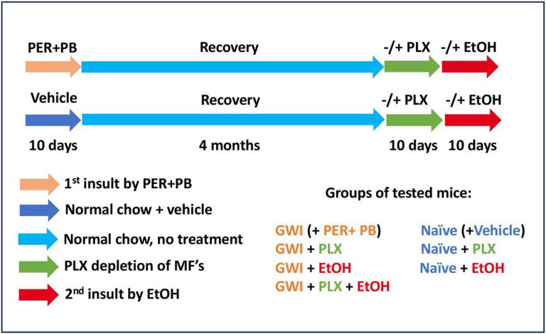
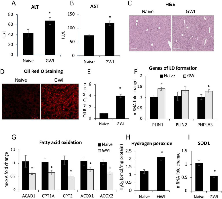
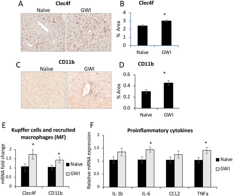
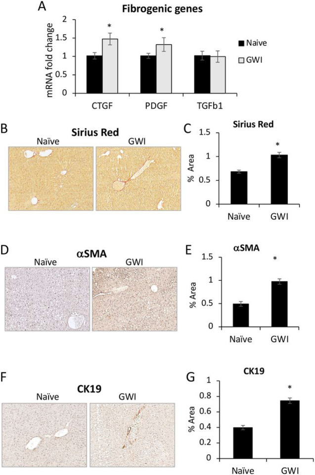
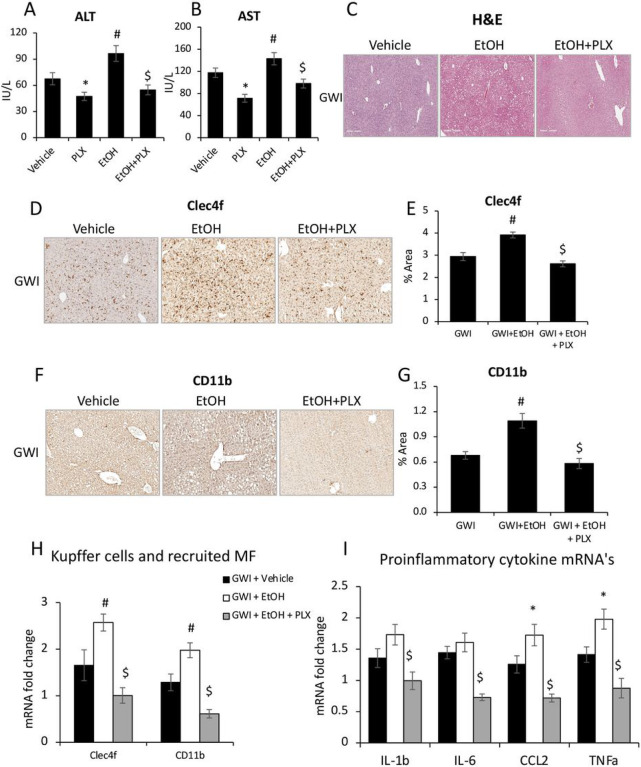
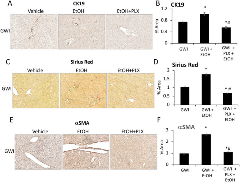
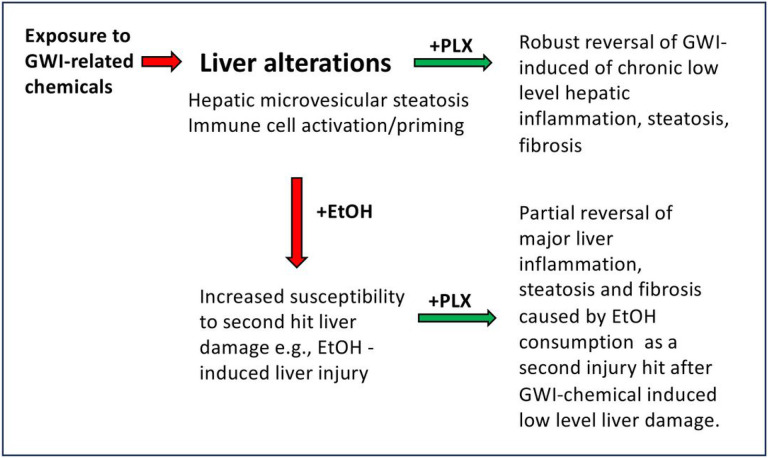
Gulf War Illness (GWI) describes a series of symptoms suffered by veterans of the Gulf war consisting of cognitive, neurological and gastrointestinal dysfunctions. Two chemicals associated with GWI are the insecticide permethrin (PER) and the nerve gas prophylactic pyridostigmine-bromide (PB). In this study we assessed the effects of PER and PB exposure on pathology and subsequent alcohol (EtOH)-induced liver injury, and the influence of a macrophage depletor, PLX3397, on EtOH-induced liver damage in PER/PB- treated mice. Male C57BL/6 mice were injected daily with vehicle or PER/PB for 10 days, followed by 4 months recovery, then treatment with PLX3397 and a chronic-plus-single-binge EtOH challenge for 10 days. PER/PB exposure resulted in the protracted increase in liver transaminases in the serum and induced chronic low-level microvesicular steatosis and inflammation in GWI vs Naïve mice up to 4 months after cessation of exposure. Furthermore, prior exposure to PER/PB also resulted in exacerbated response to EtOH-induced liver injury, with enhanced steatosis, ductular reaction and fibrosis. The enhanced EtOH-induced liver damage in GWI-mice was attenuated by strategies designed to deplete macrophages in the liver. Taken together, these data suggest that exposure to GWI-related chemicals may alter the liver's response to subsequent ethanol exposure.


Conflict of interest statement

Competing interests No conflicts of interest, financial or otherwise, are declared by the authors.

Figures

Figure 1
Figure 1
Timeline of in vivo experiments to assess the effects of GWI-related chemicals on mice exposed to PER and PB, vs Naïve mice. The additional treatments with PLX3397 inhibitor of CSF-1R in macrophages and alcohol (EtOH) were applied as shown in the diagram. Abbreviations: PER, permethrin; PB, pyridostigmine-bromide; EtOH, ethyl alcohol; PLX, PLX3397; MF, macrophages; GWI, Gulf War Illness.
Figure 2
Figure 2
Exposure of mice to GWI-related chemicals caused chronic low-grade liver damage as detected by serum transaminases and microvesicular steatosis. ALT (A) and AST (B) transaminase levels in serum samples from Naïve and GWI-mice after 4 months of recovery from exposure to PER plus PB, or vehicle. (C), Images of H&E staining of liver sections from Naïve and GWI mice. (D), Images of lipid droplets detected by Oil Red O staining of liver sections, followed by confocal microscopy. (E), Image analysis and quantification of lipid droplets as percentage of stained area, in livers from Naïve and GWI mice. (F), RT-qPCR results for expression of genes associated with lipid droplets, i.e. PLIN1, PLIN2 and PNPLA3 relative to GAPDH. (G), RT-qPCR to test the expression of genes with role in fatty acid oxidation, i.e., ACAD1, CPT1A, CPT2, ACOX1, ACOX2 in livers of Naive and GWI mice. (H), the level of hydrogen peroxide as a marker of reactive oxygen species (ROS), was determined in liver samples of GWI vs Naïve mice. (I), RT-qPCR results for SOD1 gene that encodes for superoxide dismutase, a liver enzyme with role in elimination of ROS. N=4, p<0.05, *, GWI vs Naïve mice. Scale bar, 100 mm.
Figure 3
Figure 3
Exposure of mice to GWI-related chemicals resulted in chronic low-grade hepatic inflammation. (A), IHC images of Clec4f marker of Kupffer cells. (B), Quantification of CLEC4f of Kupffer cells as percent-stained area. (C), IHC of CD11b marker of infiltrated monocyte-derived macrophages, in liver sections of Naïve and GWI-mice. (D), Image analysis results for CD11b as percent-stained area, by IHC. (E), RT-qPCR for Clec4f and CD11b in livers of Naïve and GWI mice. (F), RT-qPCR results of proinflammatory genes IL1b, IL6, CCL2 and TNFa in livers of Naïve and GWI mice. N=4, p<0.05, *, GWI vs Naïve mice. Scale bar, 100 mm.
Figure 4
Figure 4
Exposure of mice to GWI-related chemicals PER and PB, induced chronic, low-grade hepatic fibrosis in mice. (A), Changes in mRNA expression of genes with role in fibrogenesis, i.e., CTGF, PDGFb, TGFb1 in livers from Naïve and GWI mice. (B), Representative images of Sirius Red staining of liver sections. (C), Quantification of proteins that are markers of liver fibrosis, based on image analysis of Sirius Red staining in livers from Naïve and GWI mice. IHC images (D) and quantification by image analysis (E) was performed for aSMA protein that is a biomarker of activated HSC. The intrahepatic bile duct mass in livers of GWI vs Naïve mice, was measured by IHC of CK19 marker of cholangiocytes; representative images (F) and quantification (G) are shown. N=4, p<0.05, *, GWI vs Naïve mice. Scale bar, 100 mm.
Figure 5
Figure 5
The effects of PER and PB on HepG2 cells in vitro. HepG2 cells were treated with PER, PB or a combination of PER and PB as described under Methods. Changes induced by these treatments in mRNA’s encoding for PLIN2 (A), PNPLA3 (B) and SREBP1 (C), were measured using RT-qPCR. The influence of PER, PB or PER+PB on lipid accumulation in HepG2 cells, was assessed by staining of the lipids with Nile Red: images of Nile Red-labeled lipid droplets (D) and quantification of area percent staining (E) are shown. N=4, p<0.05.*PER, PB, PER+PB vs Vehicle. Scale bar, 100 mm.
Figure 6
Figure 6
The effects of ethanol (EtOH) binging on liver function in GWI mice vs Naïve mice. ALT (A) and AST (B) transaminases were measured in serum samples from Naïve and GWI mice when treated with EtOH vs negative control mice. (C), H&E staining of liver sections were observed for histopathology and steatosis. (D), Images of PLIN2 immunostaining in liver samples from Naïve/GWI mice that binged on EtOH vs mice that did not receive EtOH. (E), Image analysis data for PLIN2 IHC in the mice described for panel D. N=4, p<0.05.*GWI vs Naïve mice. #, EtOH vs no EtOH within same group of Naïve or GWI mice. $, GWI+ EtOH vs Naïve+ EtOH. Scale bar, 100 mm.
Figure 7
Figure 7
The effects of EtOH on hepatic inflammation in GWI-mice vs Naïve mice. The density of Kupffer cells was assessed by measuring Celc4f expression at mRNA (A) and protein (B, C). Changes in mRNA (D), and protein (E, F) levels of CD11b. Changes in mRNA of genes with role in inflammation (IL1b, IL6, CCL2, TNFa) were determined using RT-qPCR for livers from Naive mice (G) vs GWI mice (H) in the absence or presence of EtOH. N=4, p<0.05.*GWI vs Naïve mice. #, EtOH vs no EtOH within same group of Naïve or GWI mice. $, GWI+ EtOH vs Naïve+ EtOH. Scale bar, 100 mm.
Figure 8
Figure 8
Influence of EtOH on liver fibrosis and intrahepatic biliary duct mass in GWI-mice vs. Naïve controls. Liver sections from GWI and Naïve mice that consumed EtOH vs mice that didn’t consume EtOH, were assessed for fibrosis using Sirius Red staining of ECM proteins and aSMA IHC. (A), Representative images of Sirius Red-staining. (B), Quantification of % area stained by image analysis. Images of aSMA IHC, and quantification data based on Image analysis are shown in panels C and D, respectively. Changes in IBDM were assessed by CK19 IHC: E, representative images; (F), quantification of CK19 based on image analysis. N=4, p<0.05.*GWI vs Naïve mice. #, EtOH vs no EtOH within same group of Naïve or GWI mice. $, GWI+ EtOH vs Naïve+ EtOH. Scale bar, 100 mm.
Figure 9
Figure 9
Depletion of macrophages improved liver function, and reduced hepatic inflammation caused by EtOH consumption in GWI mice. Serum transaminases ALT (A) and AST (B), as well as images of H&E staining (C) in Naïve or GWI mice that were treated with vehicle, or PLX3397 before EtOH binging (i.e., PLX, +EtOH), are shown. IHC images of Clec4f marker of Kupffer cells, and quantification data using image analysis are illustrated in panels (D) and (E), respectively. CD11b marker of infiltrated monocyte-derived macrophages in the liver was measured by IHC (F) and quantification using image analysis (G). Changes in Clec4f and CD11b mRNA were measured in livers from GWI mice when treated with vehicle, or PLX3397 prior to EtOH (H). Relative expression of mRNA for IL-1b, IL-6, CCL2 and TNFa was measured in the same groups of mice (I). N=4, p<0.05.*PLX vs vehicle. #, EtOH vs vehicle. $, PLX+ EtOH vs EtOH. Scale bar, 100 mm.
Figure 10
Figure 10
PLX3397 mitigated the increase in ductular reaction, fibrosis and ER stress in the liver of GWI mice binging on EtOH. (A), Representative images of CK19 IHC in livers of GWI mice treated with vehicle, EtOH or EtOH+PLX. (B), Image analysis quantification of CK19 IHC. Markers of fibrosis such as collagens and other ECM proteins were stained with Sirius Red: images (C) and quantification (D) are shown. aSMA, as indicator of HSC activation, was tested by IHC; images and quantification of aSMA IHC are shown in panels (E) and (F), respectively. N=4, p<0.05.*EtOH vs vehicle. #, EtOH vs PLX+ EtOH. Scale bar, 100 mm.
Figure 11
Figure 11
Schematic representation of critical changes in the liver in mice exposed to GWI-related substances and subsequently challenged with EtOH in the absence or presence of PLX3397 inhibitor of CSF1R of macrophages. Mice that were exposed to a combination of PER and PB followed by a period of recovery, exhibited significant levels of hepatic microsteatosis and chronic low-grade inflammation and priming of the immune cells in the liver. Indeed, GWI mice that underwent chronic single binge of EtOH (a second hit on the liver), suffered increased liver damage as compared to Naïve mice with EtOH. Treatment with PLX3397, a small molecule that depletes macrophages, reduced inflammation and microsteatosis in mice exposed to GWI compared to Naïve controls. Moreover, GWI mice treated with PLX3397 prior to EtOH binging, decreased the level of steatosis, fibrosis and ductular reaction, compared to GWI mice that did not receive PLX3397.

Similar articles

References

    1. Elhaj R. & Reynolds J. M. Chemical exposures and suspected impact on Gulf War Veterans. Mil Med Res 10, 11, doi:10.1186/s40779-023-00449-9 (2023). - DOI - PMC - PubMed
    1. Engdahl B. E. et al. Brain Function in Gulf War Illness (GWI) and Associated Mental Health Comorbidities. J Neurol Neuromedicine 3, 24–34 (2018). - PMC - PubMed
    1. Engdahl B. E. et al. A Magnetoencephalographic (MEG) Study of Gulf War Illness (GWI). EBioMedicine 12, 127–132, doi:10.1016/j.ebiom.2016.08.030 (2016). - DOI - PMC - PubMed
    1. Toomey R. et al. Neuropsychological functioning of U.S. Gulf War veterans 10 years after the war. J Int Neuropsychol Soc 15, 717–729, doi:10.1017/S1355617709990294 (2009). - DOI - PubMed
    1. Kang H. K., Li B., Mahan C. M., Eisen S. A. & Engel C. C. Health of US veterans of 1991 Gulf War: a follow-up survey in 10 years. J Occup Environ Med 51, 401–410, doi:10.1097/JOM.0b013e3181a2feeb (2009). - DOI - PubMed

Publication types

LinkOut - more resources