[Preprint]. 2024 Jan 17:2024.01.15.24301339.
doi: 10.1101/2024.01.15.24301339.

Pathogenic autoantibody internalization in myositis

Affiliations

Pathogenic autoantibody internalization in myositis

Iago Pinal-Fernandez et al. medRxiv. .

Update in

  • Pathological autoantibody internalisation in myositis.
    Pinal-Fernandez I, Muñoz-Braceras S, Casal-Dominguez M, Pak K, Torres-Ruiz J, Musai J, Dell'Orso S, Naz F, Islam S, Gutierrez-Cruz G, Cano MD, Matas-Garcia A, Padrosa J, Tobias-Baraja E, Garrabou G, Aldecoa I, Espinosa G, Simeon-Aznar CP, Guillen-Del-Castillo A, Gil-Vila A, Trallero-Araguás E, Christopher-Stine L, Lloyd TE, Liewluck T, Naddaf E, Stenzel W, Greenberg SA, Grau JM, Selva-O'Callaghan A, Milisenda JC, Mammen AL. Pinal-Fernandez I, et al. Ann Rheum Dis. 2024 Oct 21;83(11):1549-1560. doi: 10.1136/ard-2024-225773. Ann Rheum Dis. 2024. PMID: 38902010 Free PMC article.

Abstract

Objectives: Myositis is a heterogeneous family of autoimmune muscle diseases. As myositis autoantibodies recognize intracellular proteins, their role in disease pathogenesis has been unclear. This study aimed to determine whether myositis autoantibodies reach their autoantigen targets within muscle cells and disrupt the normal function of these proteins.

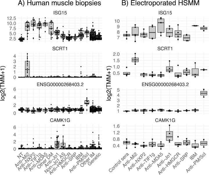
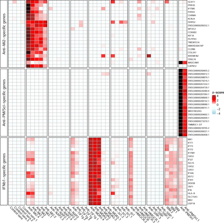
Methods: Confocal immunofluorescence microscopy was used to localize antibodies and other proteins of interest in myositis muscle biopsies. Bulk RNA sequencing was used to study the transcriptomic profiles of 668 samples from patients with myositis, disease controls, and healthy controls. Antibodies from myositis patients were introduced into cultured myoblasts by electroporation and the transcriptomic profiles of the treated myoblasts were studied by bulk RNA sequencing.

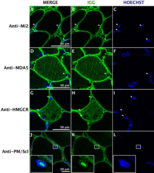
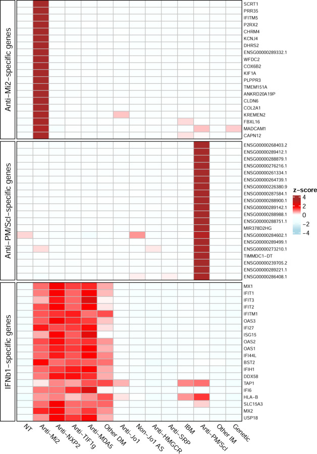
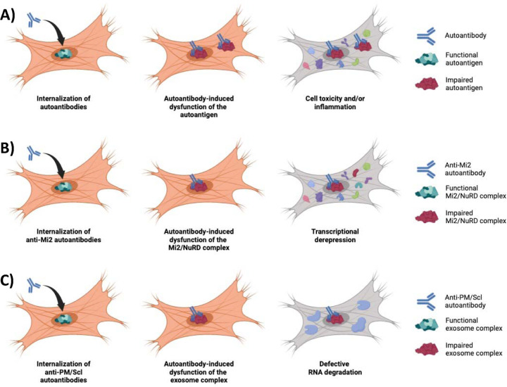
Results: In patients with myositis autoantibodies, antibodies accumulated inside myofibers in the same subcellular compartment as the autoantigen. Each autoantibody was associated with effects consistent with dysfunction of its autoantigen, such as the derepression of genes normally repressed by Mi2/NuRD in patients with anti-Mi2 autoantibodies, the accumulation of RNAs degraded by the nuclear RNA exosome complex in patients with anti-PM/Scl autoantibodies targeting this complex, and the accumulation of lipids within myofibers of anti-HMGCR-positive patients. Internalization of patient immunoglobulin into cultured myoblasts recapitulated the transcriptomic phenotypes observed in human disease, including the derepression of Mi2/NuRD-regulated genes in anti-Mi2-positive dermatomyositis and the increased expression of genes normally degraded by the nuclear RNA exosome complex in anti-PM/Scl-positive myositis.

Conclusions: In myositis, autoantibodies are internalized into muscle fibers, disrupt the biological function of their autoantigen, and mediate the pathophysiology of the disease.

Keywords: Myositis; RNA-sequencing; autoantibodies; dermatomyositis; immune-mediated necrotizing myositis.


Conflict of interest statement

Competing interests: None.

Figures

Figure 1.
Figure 1.
Immunoglobulin localization in myositis muscle. Confocal immunofluorescence of human immunoglobulin (IgG) in different autoantibody-defined types of myositis shows antibody deposition in the nuclei (white arrowheads) of muscle fibers from anti-Mi2-positive patients (A-C); the cytoplasm of anti-MDA5-positive (D-F) and anti-HMGCR-positive (G-I) patients; and the nucleoli of anti-PM/Scl-positive patients (J-L). The square box contains a higher magnification image of one nucleus, showcasing the nucleolar pattern of IgG.
Figure 2.
Figure 2.
Representative genes expressed in muscle biopsies and cultured myoblasts electroporated with antibodies from myositis patients. Muscle biopsies from all dermatomyositis subgroups (A) and cultured muscle cells with internalized antibodies from 3/5 anti-MDA5-positive patients (B) overexpressed ISG15, a representative IFNB1-stimulated gene. Anti-Mi2-positive muscle biopsies (A) and cultured cells electroporated with antibodies from anti-Mi2-positive patients (B) overexpressed SCRT1, a representative gene from the anti-Mi2-specific gene set. Anti-PM/Scl-positive muscle biopsies (A) and cultured cells electroporated with antibodies from anti-PM/Scl-positive patients (B) overexpressed ENSG00000268403.2, a representative long non-coding RNA from the anti-PM/Scl-specific gene set. Muscle biopsies from anti-synthetase patients (A) and cultured cells electroporated with antibodies from anti-Jo1 patients (B) overexpressed CAMK1G, a member of the anti-Jo1-specific gene set. NT: histologically normal muscle biopsies; DM: dermatomyositis; AS: antisynthetase syndrome; IBM: inclusion body myositis; IM: inflammatory myopathies.
Figure 3.
Figure 3.
Gene expression patterns in muscle biopsies. Standardized expression levels (Z-scores of the median) are shown for the top 20 anti-Mi2-specific genes, the top 20 anti-PM/Scl-specific genes, and the top 20 IFNB1-specific genes in muscle biopsies from patients with different types of myopathies and histologically normal muscle biopsies (NT). The set of IFNB1-specific genes was derived from RNAseq data of cultured human muscle cells treated with different type I interferons. DM: dermatomyositis; AS: antisynthetase syndrome; IBM: inclusion body myositis; IM: inflammatory myopathies.
Figure 4.
Figure 4.
Gene expression patterns in cultured human myoblasts with internalized patient antibodies. Standardized expression levels (Z-scores) of the top 20 anti-Mi2-specific genes, the top 20 anti-PM/Scl-specific genes, and the top 20 IFNB1-specific genes are shown for cultured human myoblasts electroporated with purified antibodies from individual myositis patients and healthy controls. Anti-Mi2-specific genes are overexpressed predominantly in myoblasts electroporated with antibodies from anti-Mi2-positive patients. Anti-PM/Scl-specific genes are overexpressed exclusively in myoblasts electroporated with antibodies from anti-PM/Scl-positive patients. IFNB1-specific genes are most highly expressed in myoblasts electroporated with antibodies from 3 of 5 anti-MDA5-positive patients.
Figure 5.
Figure 5.
In myositis, autoantibodies are internalized into the muscle fibers, disrupting the normal biological function of their autoantigen, which mediates the pathogenesis of the disease (A). For instance, anti-Mi2 autoantibodies (B) interfere with the Mi2/NuRD complex inducing the derepression of more than 100 genes. Similarly, anti-PM/Scl autoantibodies (C) cause a dysfunction of the nuclear RNA exosome complex, impairing the normal degradation of various types of RNA.

References

    1. Casal-Dominguez M, Pinal-Fernandez I, Pak K, et al. Performance of the 2017 European Alliance of Associations for Rheumatology/American College of Rheumatology Classification Criteria for Idiopathic Inflammatory Myopathies in Patients With Myositis-Specific Autoantibodies. Arthritis Rheumatol 2022;74(3):508–517. DOI: 10.1002/art.41964. - DOI - PMC - PubMed
    1. Allenbach Y, Arouche-Delaperche L, Preusse C, et al. Necrosis in anti-SRP(+) and anti-HMGCR(+)myopathies: Role of autoantibodies and complement. Neurology 2018. DOI: 10.1212/WNL.0000000000004923. - DOI - PubMed
    1. Arouche-Delaperche L, Allenbach Y, Amelin D, et al. Pathogenic role of anti-signal recognition protein and anti-3-Hydroxy-3-methylglutaryl-CoA reductase antibodies in necrotizing myopathies: Myofiber atrophy and impairment of muscle regeneration in necrotizing autoimmune myopathies. Ann Neurol 2017;81(4):538–548. DOI: 10.1002/ana.24902. - DOI - PubMed
    1. Bergua C, Chiavelli H, Allenbach Y, et al. In vivo pathogenicity of IgG from patients with anti-SRP or anti-HMGCR autoantibodies in immune-mediated necrotising myopathy. Ann Rheum Dis 2019;78(1):131–139. DOI: 10.1136/annrheumdis-2018-213518. - DOI - PubMed
    1. Greenberg SA, Pinkus JL, Pinkus GS, et al. Interferon-alpha/beta-mediated innate immune mechanisms in dermatomyositis. Ann Neurol 2005;57(5):664–78. DOI: 10.1002/ana.20464. - DOI - PubMed

Publication types