. 2024 Jan 18;9(4):4684-4694.
doi: 10.1021/acsomega.3c07947. eCollection 2024 Jan 30.

Allosteric Signal within the Receptor-Binding Domain of the SARS-CoV-2 Spike Protein Mediated by a Class 3 Monoclonal Antibody Revealed through Molecular Dynamics Simulations and Protein Residue Networks

Affiliations

Allosteric Signal within the Receptor-Binding Domain of the SARS-CoV-2 Spike Protein Mediated by a Class 3 Monoclonal Antibody Revealed through Molecular Dynamics Simulations and Protein Residue Networks

Patamalai Boonserm et al. ACS Omega. .

Abstract

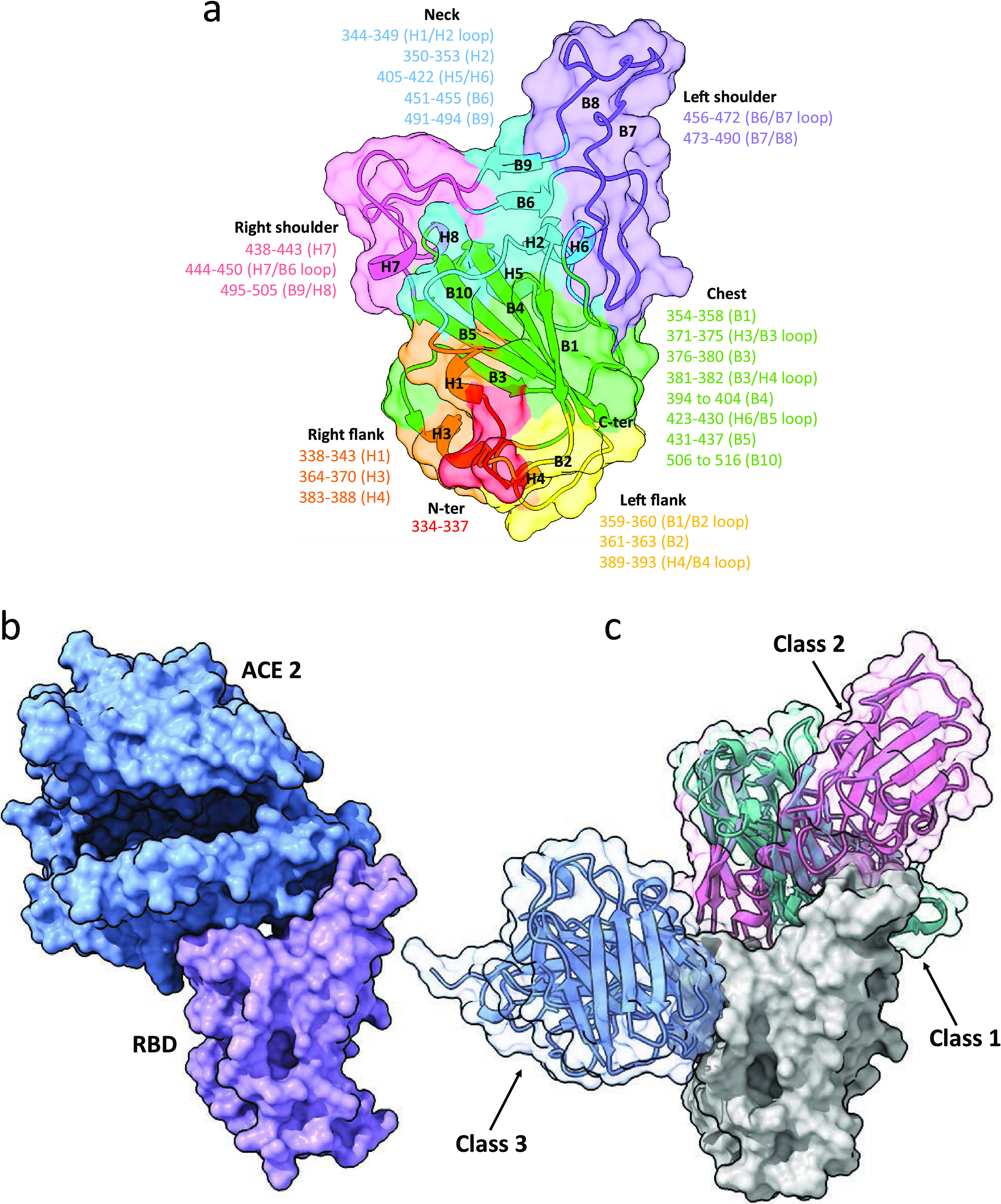
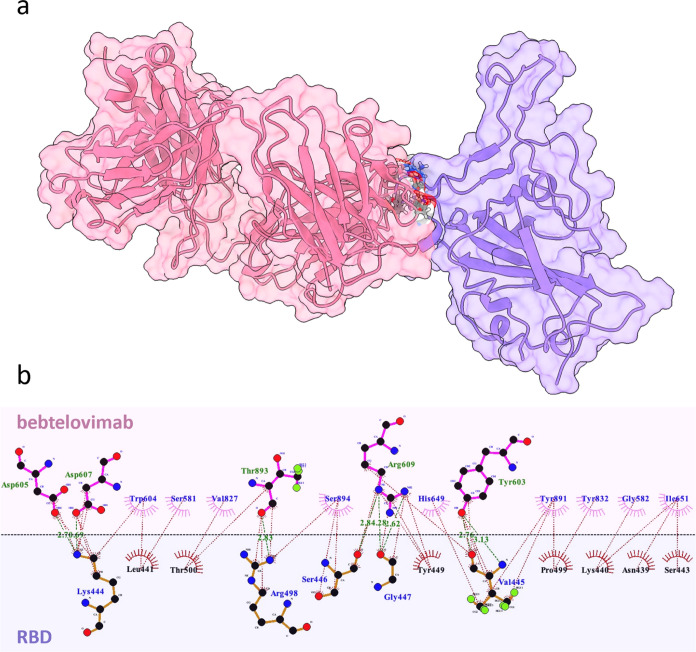
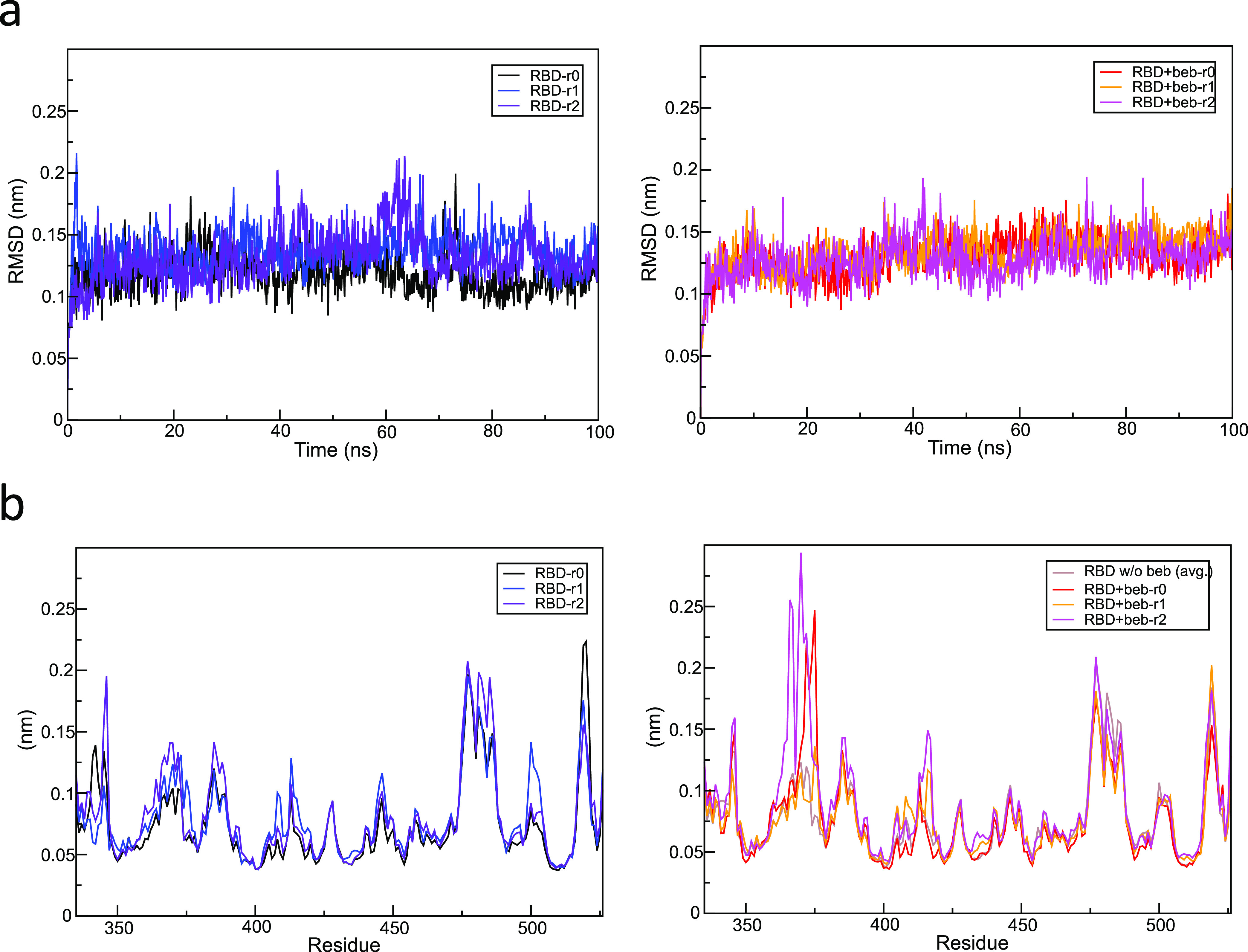
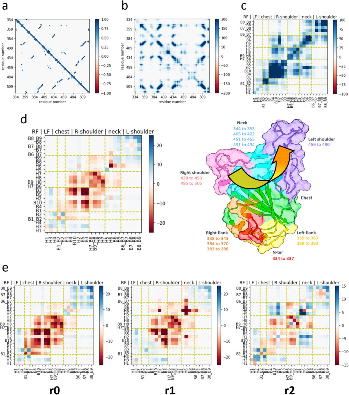
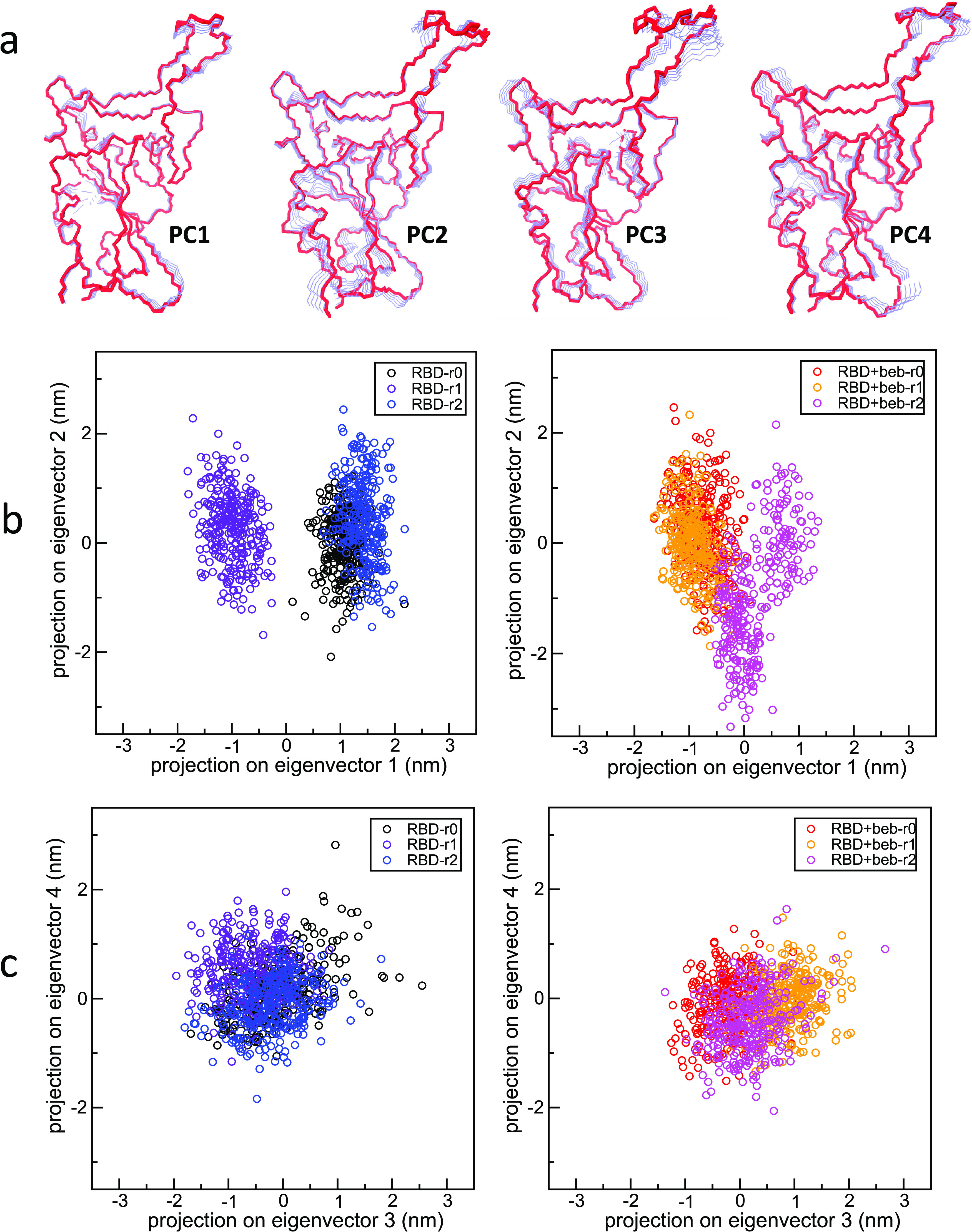
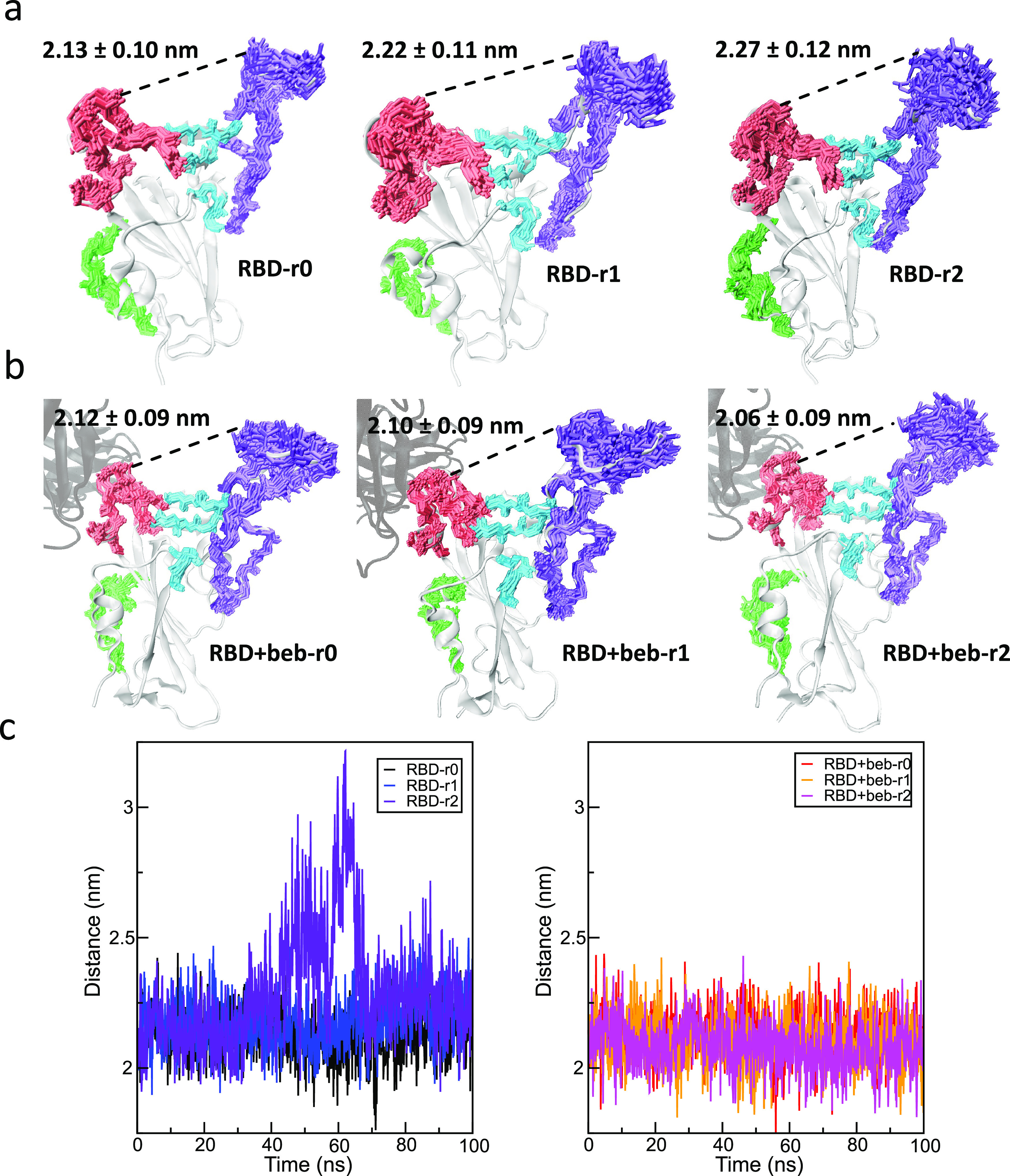
This study investigated the allosteric action within the receptor-binding domain (RBD) of the SARS-CoV-2 spike protein caused by class 3 monoclonal antibody (mAb) binding. As the emergence of SARS-CoV-2 variants has raised concerns about the effectiveness of treatments by antibodies, targeting the highly conserved class 3 epitopes has become an alternative strategy of antibody design. Simulations of explicitly solvated RBD of the BA.2.75 omicron subvariants were carried out both in the presence and in the absence of bebtelovimab, as a model example of class 3 monoclonal antibodies against the RBD of the SARS-CoV-2 spike protein. The comparative analysis showed that bebtelovimab's binding on two α helices at the epitope region disrupted the nearby interaction network, which triggered a denser interaction network formation on the opposite side of the receptor-binding motif (RBM) region and resulted in a "close" conformation that could prevent the ACE2 binding. A better understanding of this allosteric action could lead to the development of alternative mAbs for further variants of concern. In terms of computational techniques, the communicability matrix could serve as a tool to visualize the effects of allostery, as the pairs of amino acids or secondary structures with high communicability could pinpoint the possible sites to transfer the allosteric signal. Additionally, the communicability gain/loss matrix could help elucidate the consequences of allosteric actions, which could be employed along with other allostery quantification techniques in some previous studies.


Conflict of interest statement

The authors declare no competing financial interest.

Figures

Figure 1
Figure 1
(a) Nomenclatures for different regions and secondary structures of a receptor-binding domain (RBD) of SARS-CoV-2 spike proteins, (b) conformation of an RBD/ACE2 complex obtained from the protein databank (PDB ID: 6M0J), and (c) an RBD domain with superimposed binding configurations of class 1–3 antibodies at different epitopes.
Figure 2
Figure 2
(a) Representative conformation of an RBD (purple)/bebtelovimab (pink) complex obtained from three 100 ns explicitly solvated atomistic molecular dynamics (MD) simulations and (b) interaction network between bebtelovimab and an RBD at the binding interface.
Figure 3
Figure 3
(a) Root-mean-square deviation (RMSD) relative to starting structures along all 100 ns MD trajectory replicas of the RBD in the absence (left) and in the presence (right) of bebtelovimab binding and (b) per-residue root-mean-square fluctuation (RMSF) of three RBD replicas in the absence (left) and in the presence (right) of bebtelovimab binding compared with the average RMSF of all replicas without bebtelovimab.
Figure 4
Figure 4
(a) Time-averaged adjacency matrix of an RBD domain without bebtelovimab over the last 30 ns of all three MD replicas; (b) time-averaged communicability matrix of an RBD domain without bebtelovimab; (c) communicability matrix of an RBD domain averaged over the last 30 ns of all three MD replicas and over the residues within secondary structures; (d) matrix of communicability gain/loss defined by the difference in communicability within the RBD structure without bebtelovimab binding and the RBD structure with bebtelovimab binding, averaged over three replicas; and (e) communicability gain/loss defined by the difference in communicability within the RBD structure without bebtelovimab binding and the RBD structure with bebtelovimab binding for each replica. The yellow arrow illustrates the direction of allosteric signal transfer.
Figure 5
Figure 5
(a) First four essential (PCA) modes extracted from the combined RBD trajectory (the positive extremum is presented in red color), (b) projection of RBD trajectories in the absence (left) and in the presence (right) of bebtelovimab onto the first and second PCA modes, and (c) projection of RBD trajectories in the absence (left) and in the presence (right) of bebtelovimab onto the third and fourth PCA modes.
Figure 6
Figure 6
(a, b) Superimposed conformational snapshots of the right shoulder (pink), neck (light blue), and left shoulder (purple) regions captured every 3 ns between 70 and 100 ns for all MD replicas of RBD (a) without bebtelovimab binding and (b) with bebtelovimab binding. The yellow arrow illustrates the direction of allosteric signal transfer due to bebtelovimab binding. Solid lines were drawn between residues 446 and 484. (c) Minimum distance between residues 446 and 484 (left) without bebtelovimab binding and (right) with bebtelovimab binding.

Similar articles

Cited by

References

    1. Kim S.-M.; Kim E. H.; Casel M. A. B.; et al. SARS-CoV-2 variants with NSP12 P323L/G671S mutations display enhanced virus replication in ferret upper airways and higher transmissibility. Cell Rep. 2023, 42 (9), 11307710.1016/j.celrep.2023.113077. - DOI - PMC - PubMed
    1. Zhou H.; Møhlenberg M.; Thakor J. C.; et al. Sensitivity to Vaccines, Therapeutic Antibodies, and Viral Entry Inhibitors and Advances To Counter the SARS-CoV-2 Omicron Variant. Clin. Microbiol. Rev. 2022, 35 (3), e00014-2210.1128/cmr.00014-22. - DOI - PMC - PubMed
    1. Jackson C. B.; Farzan M.; Chen B.; et al. Mechanisms of SARS-CoV-2 entry into cells. Nat. Rev. Mol. Cell Biol. 2022, 23 (1), 3–20. 10.1038/s41580-021-00418-x. - DOI - PMC - PubMed
    1. Fehr A. R.; Perlman S. Coronaviruses: an overview of their replication and pathogenesis. Methods Mol. Biol. 2015, 1282, 1–23. 10.1007/978-1-4939-2438-7_1. - DOI - PMC - PubMed
    1. Basavarajappa S. C.; Liu A. R.; Bruchez A.; et al. Trimeric receptor-binding domain of SARS-CoV-2 acts as a potent inhibitor of ACE2 receptor-mediated viral entry. iScience 2022, 25 (8), 10471610.1016/j.isci.2022.104716. - DOI - PMC - PubMed

LinkOut - more resources