. 2024 Jan 18;9(4):4920-4930.
doi: 10.1021/acsomega.3c08632. eCollection 2024 Jan 30.

A Salt Bridge and Disulfide Bond within the Lassa Virus Fusion Domain Are Required for the Initiation of Membrane Fusion

Affiliations

A Salt Bridge and Disulfide Bond within the Lassa Virus Fusion Domain Are Required for the Initiation of Membrane Fusion

Hallie N Pennington et al. ACS Omega. .

Abstract

Infection with Lassa virus (LASV), an Old-World arenavirus that is endemic to West Africa, causes Lassa fever, a lethal hemorrhagic fever. Delivery of LASV's genetic material into the host cell is an integral component of its lifecycle. This is accomplished via membrane fusion, a process initiated by a hydrophobic sequence known as the fusion domain (FD). The LASV FD (G260-N295) consists of two structurally distinct regions: an N-terminal fusion peptide (FP: G260-T274) and an internal fusion loop (FL: C279-N295) that is connected by a short linker region (P275-Y278). However, the molecular mechanisms behind how the LASV FD initiates fusion remain unclear. Here, we demonstrate that the LASV FD adopts a fusogenic, helical conformation at a pH akin to that of the lysosomal compartment. Additionally, we identified a conserved disulfide bond (C279 and C292) and salt bridge (R282 and E289) within the FL that are pertinent to fusion. We found that the disulfide bond must be present so that the FD can bind to the lipid bilayer and subsequently initiate fusion. Moreover, the salt bridge is essential for the secondary structure of the FD such that it can associate with the lipid bilayer in the proper orientation for full functionality. In conclusion, our findings indicate that the LASV FD preferentially initiates fusion at a pH akin to that of the lysosome through a mechanism that requires a conserved salt bridge and, to a lesser extent, an intact disulfide bond within the internal FL.


Conflict of interest statement

The authors declare no competing financial interest.

Figures

Figure 1
Figure 1
Several regions within the LASV glycoprotein complex (GPC) that have functional relevance to membrane fusion. The GPC is cleaved by SPase at position 58, yielding the stable signal peptide (SSP), which has a myristoylation motif (myr) at position 2, and receptor binding subunit (glycoprotein 1, (GP1)). Following cleavage by SKI-1/S1P at position 259, the fusion subunit (glycoprotein 2, (GP2)) is separated from GP1, releasing a fusion domain (FD, red) at the N-terminus that is composed of two structurally distinct regions: a fusion peptide (FP, yellow) and internal fusion loop (FL, orange) conjoined by a short linker region (dotted line). A structural alignment with two of the most common arenaviruses afflicting humans revealed that the FL contains a conserved disulfide bond (blue, solid line) and salt bridge (purple, dashed line). Residues with full conservation are indicated by an asterisk (*), whereas those with strong and weak similarities are denoted by a semicolon (:) and period (.), respectively. Abbreviations: LCMV, lymphocytic choriomeningitis virus; LUJOV, Lujo virus; HR1, heptad repeat 1; HR2, heptad repeat 2; TM, transmembrane domain.
Figure 2
Figure 2
Function and secondary structure of the LASV FD have a large pH dependence. [A] Positive correlation between pH and fusion with optimal fusion occurring at a pH of 4.0, which is akin to the lysosome (n ≥ 6). Data normalized to pH 4. [B] Global secondary structure is dependent on environmental pH with a helical structure only adopted at pH 4.
Figure 3
Figure 3
A conserved disulfide bond is required for efficient function of the LASV FD, but not for the global secondary structure. [A] Disruption of the disulfide bond reduced fusion (n ≥ 15). Data normalized to WT. Student’s t-Test assuming unequal variances used to calculate P- value; *** = P < 0.001. [B] Global secondary structure remained relatively unchanged under reducing conditions.
Figure 4
Figure 4
Interruption of the disulfide bond resulted in a decreased binding affinity of the LASV FD. [A] WT and [B] WT with 1 mM TCEP. Both ITC experiments were conducted in 10 mM NaOAc, 100 mM NaCl, pH 4.0 with vesicles titrated into the protein. Dissociation constants (Kd) are displayed on the respective isotherm.
Figure 5
Figure 5
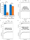
High salt concentrations impair the functionality of the LASV FD, but not the SARS-CoV-2 FD. [A] Introduction of 500 mM NaCl to the buffer system significantly reduced fusion for the LASV FD, but not SARS-CoV-2 FD (n ≥ 18). Data normalized to 100 mM NaCl for each FD. Student’s t-Test assuming unequal variances used to calculate P-value; ** = P < 0.01, *** = P < 0.001. [B] LASV FD with 500 mM NaCl; [C] SARS-CoV-2 FD; and [D] SARS-CoV-2 FD with 500 mM NaCl. All ITC experiments conducted in 10 mM NaOAc, 100 mM NaCl or 500 mM NaCl at pH 5.0 (SARS-CoV-2) or pH 4.0 (LASV), with vesicles titrated into the protein. Dissociation constants (Kd) are displayed on the respective isotherm.
Figure 6
Figure 6
An identified salt bridge is critical to the overall structure and function of the LASV FD. [A] Under physiological salt conditions (100 mM NaCl), all salt bridge mutants (R282E, red; E289R, green; R282E/E289R, purple) had decreased fusion in comparison to WT (blue) (n ≥ 38). All lipid mixing normalized to WT, pH 4. Student’s t test assuming unequal variances used to calculate P-value; n.s. = not significant, ** = P < 0.01, and *** = P < 0.001. [B] Mutation of any salt bridge residue altered the secondary structure.

References

    1. Mehand M. S.; Al-Shorbaji F.; Millett P.; Murgue B. The WHO R&D Blueprint: 2018 review of emerging infectious diseases requiring urgent research and development efforts. Antiviral Res. 2018, 159, 63–67. 10.1016/j.antiviral.2018.09.009. - DOI - PMC - PubMed
    1. Richmond J. K.; Baglole D. J. Lassa fever: epidemiology, clinical features, and social consequences. British Medical Journal 2003, 327 (7426), 1271–1275. 10.1136/bmj.327.7426.1271. - DOI - PMC - PubMed
    1. McCormick J. B.; Webb P. A.; Krebs J. W.; Johnson K. M.; Smith E. S. A Prospective Study of the Epidemiology and Ecology of Lassa Fever. J. Infect. Diseases 1987, 135 (3), 437–444. 10.1093/infdis/155.3.437. - DOI - PubMed
    1. Lecompte E.; Fichet-Calvet E.; Daffis S.; Koulémou K.; Sylla O.; Kourouma F.; Doré A.; Soropogui B.; Aniskin V.; Allali B.; et al. Mastomys natalensis and Lassa fever, West Africa. Emerging Infectious Diseases 2006, 12 (12), 1971–1974. 10.3201/eid1212.060812. - DOI - PMC - PubMed
    1. Safronetz D.; Lopez J. E.; Sogoba N.; Traore’ S. F.; Raffel S. J.; Fischer E. R.; Ebihara H.; Branco L.; Garry R. F.; Schwan T. G.; Feldmann H.; et al. Detection of Lassa virus. Mali. Emerging Infect. Dis. 2010, 16 (7), 1123–1126. 10.3201/eid1607.100146. - DOI - PMC - PubMed

LinkOut - more resources