Case Reports
. 2023 Nov 18;6(1):107-113.
doi: 10.1016/j.jhsg.2023.10.004. eCollection 2024 Jan.

Open Dislocation of the Scaphoid With an Associated Hamate Fracture and Fourth Metacarpal Fracture

Affiliations
Case Reports

Open Dislocation of the Scaphoid With an Associated Hamate Fracture and Fourth Metacarpal Fracture

Cay Mierisch et al. J Hand Surg Glob Online. .

Abstract

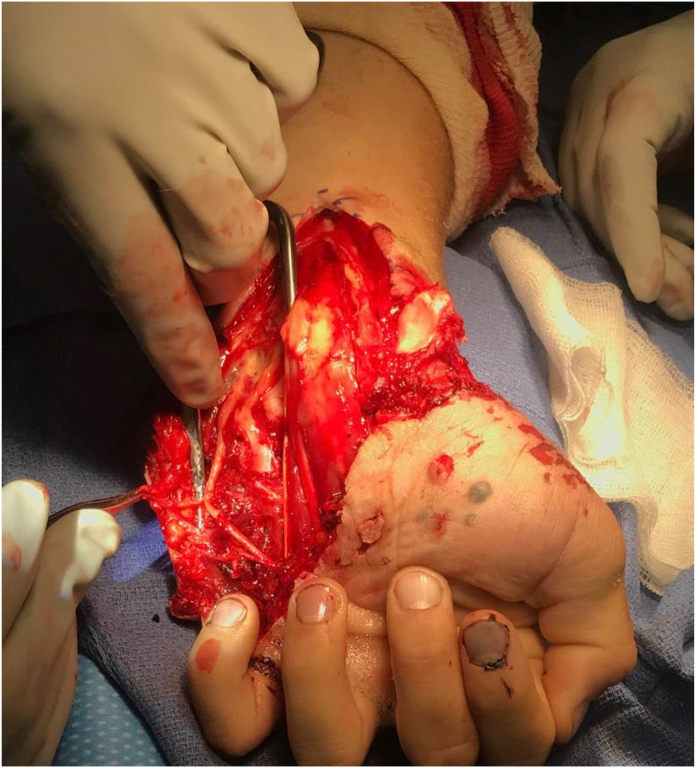
Scaphoid dislocation represents a rare injury with only a few case reports and limited case series reported in the literature. The majority of scaphoid dislocations result from a high-energy trauma causing hyperextension and ulnar deviation of the wrist. The severity of a scaphoid dislocation depends on the degree of periscaphoid ligamentous injury as well as the presence of concomitant injuries, such as axial carpal dissociation. The most common complication after a scaphoid dislocation is scapholunate dissociation, which emphasizes the importance of scapholunate ligament repair/reconstruction in these cases. We report a case of an open scaphoid dislocation with the associated injuries of a hamate fracture and fourth metacarpal fracture treated with an open reduction of the scaphoid, open ligamentous repair and augmentation of the involved carpal ligaments, and open reduction internal fixation of both the hamate and the fourth metacarpal fractures.

Keywords: Fourth metacarpal fracture; Hamate fracture; Open; Scaphoid dislocation; Volar.


Figures

Figure 1
Figure 1
An approximately 20-cm laceration from the volar radial aspect of the proximal wrist extending distally to the volar ulnar aspect of the base of the little finger. The dislocated scaphoid is directly visualized within the laceration to be distally tethered by soft tissue attachments.
Figure 2
Figure 2
Three radiographic views of the left wrist. A Posterior-anterior view. B Oblique view. C Lateral view. These images demonstrate a palmar–radial scaphoid dislocation, a hamate body fracture at the base of the hook of hamate, and a fourth metacarpal midshaft fracture.
Figure 3
Figure 3
Intraoperative image depicting the surgical release of the ulnar nerve and its branches. The scaphoid is also seen dislocated.
Figure 4
Figure 4
Two fluoroscopic views of the left wrist. A Posterior-anterior view. B Lateral view. These images demonstrate reduction and fixation of the hamate fracture with two interfragmentary screws as well as a reduced scaphoid into the scaphoid fossa.
Figure 5
Figure 5
Two fluoroscopic views of the left hand. A Posterior-anterior view. B Lateral view. These images demonstrate reduction and fixation of the metacarpal fracture with a 2-mm plate. The Nano Corkscrew FT can also be visualized in the triquetrum. The scapholunate interval is optimally reduced.
Figure 6
Figure 6
Two radiographic views of the left wrist. A Posterior-anterior view. B Lateral view. These images demonstrate optimal reduction and fixation of the patient’s multiple injuries. A vessel loop used to aid in superficial suture removal can be visualized.
Figure 7
Figure 7
The sequence of ligamentous failure in chronological order. A A normal, intact wrist. B A torn scapholunate ligament (SL) and torn radioscapholunate ligament (RSL) resulting in scapholunate dissociation. C An additionally torn radioscaphocapitate ligament (RSC) resulting in a partial scaphoid dislocation. D An additionally torn scaphotrapezial ligament (ST) resulting in a total scaphoid dislocation.
Figure 8
Figure 8
A comparison of two wrists. A A normal wrist. B A wrist with axial carpal dissociation indicated by the disruption of the capitohamate and middle–ring metacarpal base joints because of proximal migration of the radial carpus, inducing a scaphoid dislocation. The red arrow and dotted line represent the axial force transmission causing this injury pattern.

References

    1. Ely L.W. VI. Dislocation of the carpal scaphoid. Ann Surg. 1903;38(1):97–98.1. - PMC - PubMed
    1. Amundsen A., Bishop S.N., Moran S.L. Isolated scaphoid dislocation: a case report and review of the literature. J Wrist Surg. 2020;9(5):431–439. - PMC - PubMed
    1. Chloros G.D., Themistocleous G.S., Zagoreos N.P., et al. Isolated dislocation of the scaphoid. Arch Orthop Trauma Surg. 2006;126(3):197–203. - PubMed
    1. Amamilo S.C., Uppal R., Samuel A.W. Isolated dislocation of carpal scaphoid. J Hand Surg Br. 1985;10(3):385–388. - PubMed
    1. Thompson T.C., Campbell R.D., Arnold W.D. Primary and secondary dislocation of the scaphoid bone. J Bone Joint Surg Br. 1964;46(1):73–82. - PubMed

Publication types

LinkOut - more resources