

Electroencephalogram findings in 10 patients with post-stroke epilepsy: A retrospective study
- PMID: 38313653
- PMCID: PMC10835697
- DOI: 10.12998/wjcc.v12.i2.249
Electroencephalogram findings in 10 patients with post-stroke epilepsy: A retrospective study
Abstract
Background: Post-stroke epilepsy is a common and easily overlooked complication of acute cerebrovascular disease. Long-term seizures can seriously affect the prognosis and quality of life of patients. Electroencephalogram (EEG) is the simplest way to diagnose epilepsy, and plays an important role in predicting seizures and guiding medication.
Aim: To explore the EEG characteristics of patients with post-stroke epilepsy and improve the detection rate of inter-seizure epileptiform discharges.
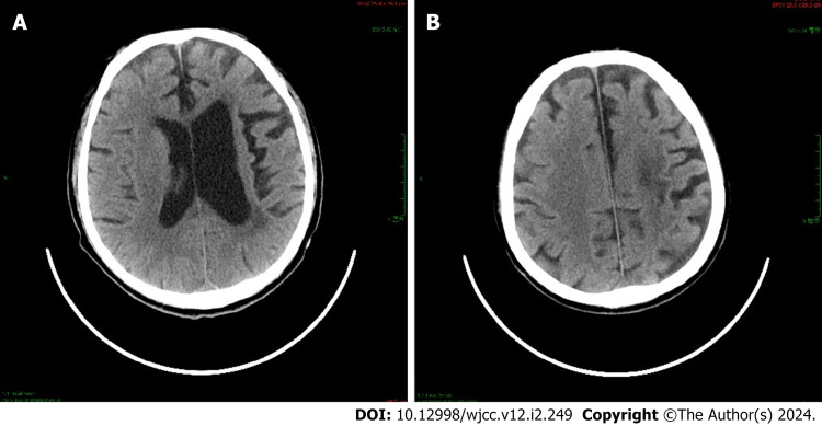
Methods: From January 2017 to June 2020, 10 patients with post-stroke epilepsy in our hospital were included. The clinical, imaging, and EEG characteristics were collected. The stroke location, seizure type, and ictal and interictal EEG manifestations of the patients with post-stroke epilepsy were then retrospectively analyzed.
Results: In all 10 patients, epileptiform waves occurred in the side opposite to the stroke lesion during the interictal stage; these manifested as sharp wave, sharp-wave complex, or spike discharges in the anterior head lead of the side opposite to the lesion.
Conclusion: In EEG, epileptiform waves can occur in the side opposite to the stroke lesion in patients with post-stroke epilepsy.
Keywords: Electroencephalogram; Post-stroke epilepsy; Seizure; Slow wave; Stroke.
©The Author(s) 2024. Published by Baishideng Publishing Group Inc. All rights reserved.
Conflict of interest statement
Conflict-of-interest statement: All authors declare that there is no conflict of interest.
Figures


References
-
- Labovitz DL, Hauser WA, Sacco RL. Prevalence and predictors of early seizure and status epilepticus after first stroke. Neurology. 2001;57:200–206. - PubMed
-
- Tanaka T, Ihara M. Post-stroke epilepsy. Neurochem Int. 2017;107:219–228. - PubMed
-
- Bentes C, Franco AC, Peralta AR, Viana P, Martins H, Morgado C, Casimiro C, Fonseca C, Geraldes R, Canhão P, Pinho E Melo T, Paiva T, Ferro JM. Epilepsia partialis continua after an anterior circulation ischaemic stroke. Eur J Neurol. 2017;24:929–934. - PubMed
-
- Arntz RM, Maaijwee NA, Rutten-Jacobs LC, Schoonderwaldt HC, Dorresteijn LD, van Dijk EJ, de Leeuw FE. Epilepsy after TIA or stroke in young patients impairs long-term functional outcome: the FUTURE Study. Neurology. 2013;81:1907–1913. - PubMed
LinkOut - more resources
Full Text Sources
