. 2024 Jan 9;4(1):100142.
doi: 10.1016/j.bpr.2024.100142. eCollection 2024 Mar 13.

Machine learning topological defects in confluent tissues

Affiliations

Machine learning topological defects in confluent tissues

Andrew Killeen et al. Biophys Rep (N Y). .

Abstract

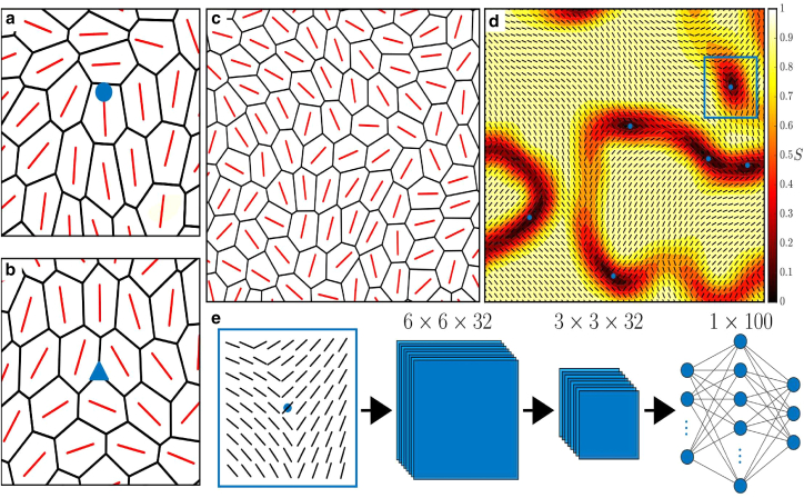
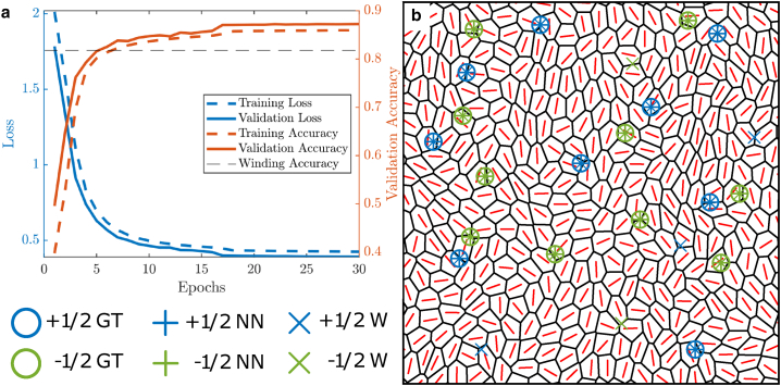
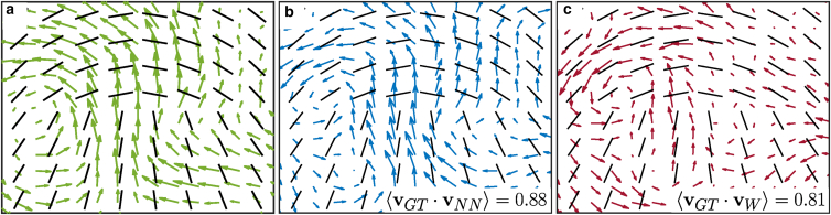
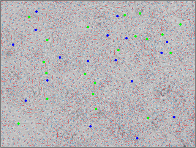
Active nematics is an emerging paradigm for characterizing biological systems. One aspect of particularly intense focus is the role active nematic defects play in these systems, as they have been found to mediate a growing number of biological processes. Accurately detecting and classifying these defects in biological systems is, therefore, of vital importance to improving our understanding of such processes. While robust methods for defect detection exist for systems of elongated constituents, other systems, such as epithelial layers, are not well suited to such methods. Here, we address this problem by developing a convolutional neural network to detect and classify nematic defects in confluent cell layers. Crucially, our method is readily implementable on experimental images of cell layers and is specifically designed to be suitable for cells that are not rod shaped, which we demonstrate by detecting defects on experimental data using the trained model. We show that our machine learning model outperforms current defect detection techniques and that this manifests itself in our method as requiring less data to accurately capture defect properties. This could drastically improve the accuracy of experimental data interpretation while also reducing costs, advancing the study of nematic defects in biological systems.


Conflict of interest statement

The authors declare no competing interests.

Figures

Figure 1
Figure 1
Topological defect identification and classification procedure. Examples of (a) comet-shaped +1/2 and (b) trefoil-shaped 1/2 defects in a confluent cell layer with the orientation of the long axis of each cell plotted in red. (c) Example of an active vertex model configuration. We find the x and y coordinates of each cell’s center of mass, and the orientation of each cell’s long axis is then plotted at these points. (d) This information is then interpolated to a finer grid to form the nematic field of the system, where the average local scalar nematic order parameter S can be calculated at each grid point using a sliding window. Areas of low nematic order (Sth<0.15) are identified as possible defect regions, and the centers of mass of these regions are identified (blue dots). The nematic field around these points is then cropped to form a region of interest (ROI) (blue box). (e) These ROIs are then input into a machine learning model, which classifies them as a +1/2 defect, a 1/2 defect, or not a defect.
Figure 2
Figure 2
Machine learning model outperforms winding number classifier. (a) The training and validation loss and accuracy of the neural network as it is trained. Mean value of 50 realizations is plotted. The black dashed line represents the accuracy of the winding number classification on the training data (0.812). (b) An example domain with defects detected using each method: our ground truth, neural network, and winding number.
Figure 3
Figure 3
Less data are needed to characterize defect properties. Average velocity fields around +1/2 defects for (a) manually labeled defects, (b) defects detected using the neural network, and (c) defects detected using the winding number. The single-point correlation function (vGT·v) between the ground-truth field and the neural network and winding number fields is also shown.
Figure 4
Figure 4
Machine learning model detects defects on experimental data. A representative example of nematic defects detected on a wild-type MDCK cell monolayer, with +1/2 defects in blue and 1/2 in green. As well as the bright-field image of the cell layer, we also plot the long axis of each cell in red and the center of mass as a light blue circle.

Similar articles

References

    1. Poujade M., Grasland-Mongrain E., et al. Silberzan P. Collective migration of an epithelial monolayer in response to a model wound. Proc. Natl. Acad. Sci. USA. 2007;104:15988–15993. - PMC - PubMed
    1. Friedl P., Gilmour D. Collective cell migration in morphogenesis, regeneration and cancer. Nat. Rev. Mol. Cell Biol. 2009;10:445–457. - PubMed
    1. Weijer C.J. Collective cell migration in development. J. Cell Sci. 2009;122:3215–3223. - PubMed
    1. Alert R., Trepat X. Physical Models of Collective Cell Migration. Annu. Rev. Condens. Matter Phys. 2020;11:77–101.
    1. Marchetti M.C., Joanny J.F., et al. Simha R.A. Hydrodynamics of soft active matter. Rev. Mod. Phys. 2013;85:1143–1189.

LinkOut - more resources