. 2024 Jan 11;35(1):102115.
doi: 10.1016/j.omtn.2024.102115. eCollection 2024 Mar 12.

Engineering lentivirus envelope VSV-G for liver targeted delivery of IDOL-shRNA to ameliorate hypercholesterolemia and atherosclerosis

Affiliations

Engineering lentivirus envelope VSV-G for liver targeted delivery of IDOL-shRNA to ameliorate hypercholesterolemia and atherosclerosis

Wei Wang et al. Mol Ther Nucleic Acids. .

Abstract

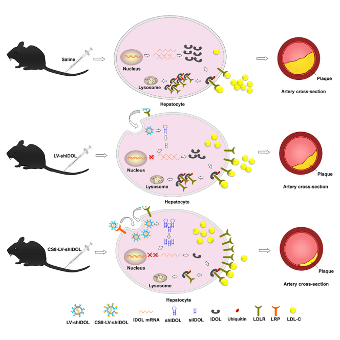
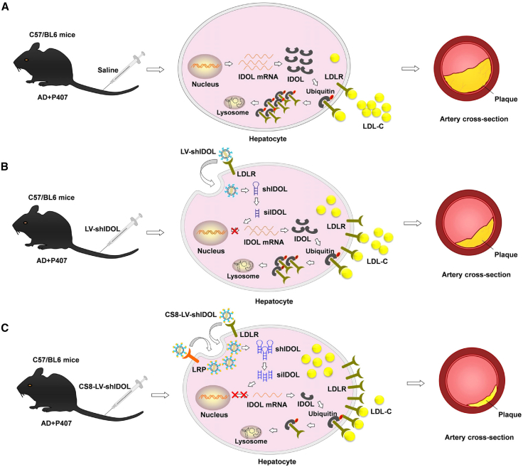
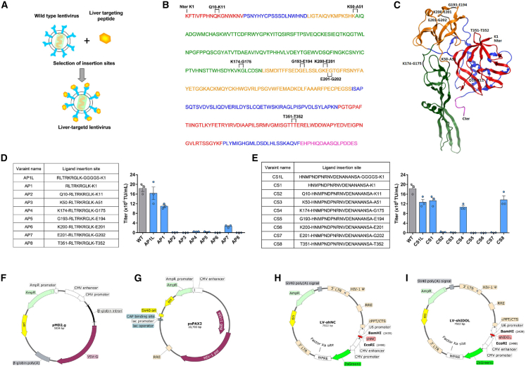
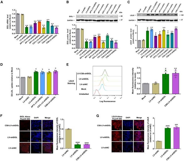
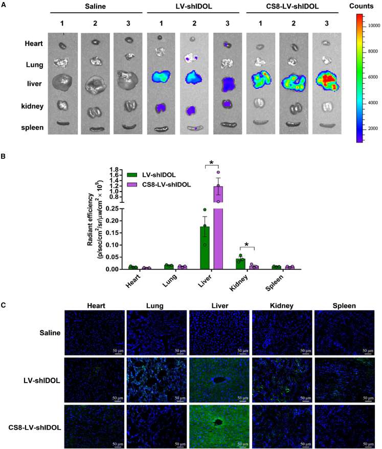
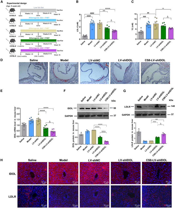
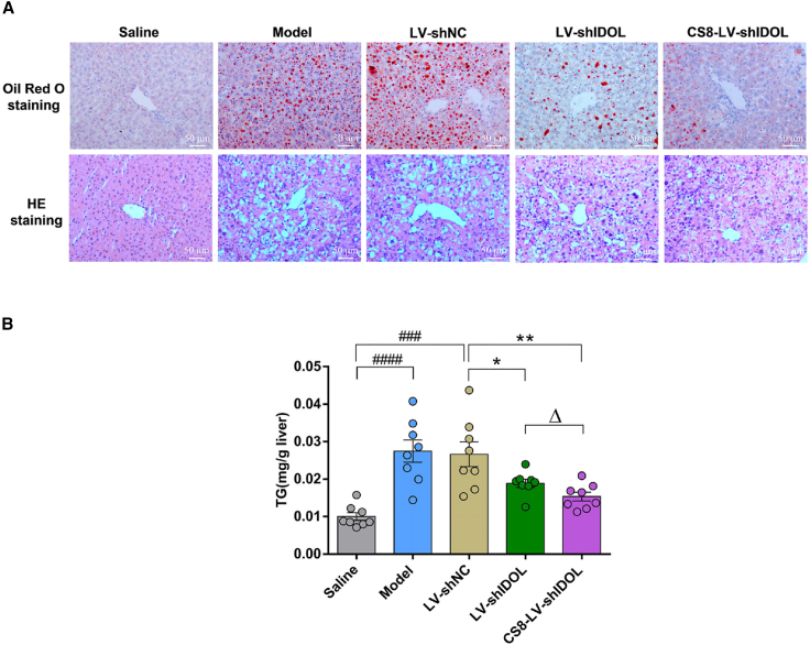
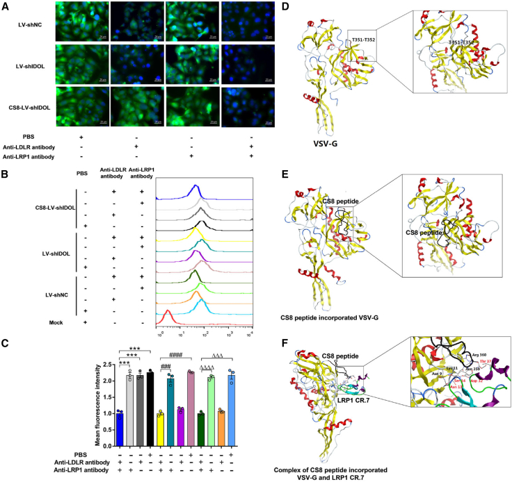
Lentiviral vectors (LVs) have been widely used as a tool for gene therapies. However, tissue-selective transduction after systemic delivery remains a challenge. Inducible degrader of low-density lipoprotein receptor is an attractive target for treating hypercholesterolemia. Here, a liver-targeted LV, CS8-LV-shIDOL, is developed by incorporating a hepatocyte-targeted peptide derived from circumsporozoite protein (CSP) into the lentivirus envelope for liver-targeted delivery of IDOL-shRNA (short hairpin RNA) to alleviate hypercholesterolemia. Tail-vein injection of CS8-LV-shIDOL results in extremely high accumulation in liver and nearly undetectable levels in other organs in mice. In addition, it shows superior therapeutic efficacy in lowering serum low-density lipoprotein cholesterol (LDL-C) and reducing atherosclerotic lesions over unmodified LV-shIDOL in hyperlipidemic mice. Mechanically, the envelope-engineered CS8-LV-shIDOL can enter liver cells via low-density lipoprotein receptor-related protein (LRP). Thus, this study provides a novel approach for liver-targeted delivery of IDOL-shRNA to treat hypercholesterolemia by using an envelope-engineered LV, and this delivery system has great potential for liver-targeted transgene therapy.

Keywords: IDOL; MT: Oligonucleotides: Therapies and Applications; gene therapy; lentivirus; liver-targeted; siRNA; small interfering RNA.


Conflict of interest statement

The authors declare no competing interests.

Figures

None
Graphical abstract
Figure 1
Figure 1
Schematic diagram illustrating the mechanism of liver-targeted lentiviral vector CS8-LV-shIDOL treating atherosclerosis in C57/BL6 mice (A) P407-induced hyperlipidemic mice fed an atherogenic diet (AD) treated with saline. IDOL binds to and promotes ubiquitination of the intracellular tail of the LDLR, resulting in lysosomal degradation of the receptor. The aortas of P407-induced atherogenic mice show pathological lesions that are thickened. (B) P407-induced hyperlipidemic mice fed an AD treated with wild-type lentiviral vector LV-shIDOL. Lentiviral vector mediated shRNA knockdown of IDOL up-regulates LDLR and lowers circulating LDL levels. The lesions of the aorta in LV-shIDOL-treated atherogenic mice are milder than those in untreated atherogenic mice. (C) P407-induced hyperlipidemic mice fed an AD treated with liver-targeted CS8-LV-shIDOL. Compared with LV-shIDOL, liver-targeted CS8-LV-shIDOL has higher potency in inhibiting hepatic IDOL in vivo, resulting in higher LDLR levels and lower serum LDL levels. The lesions of the aorta in CS8-LV-shIDOL-treated atherogenic mice are milder than those in LV-shIDOL-treated atherogenic mice.
Figure 2
Figure 2
Analysis on viable insertion sites on VSV-G for displaying hepatocyte-targeting peptides (A) Elements of secondary structure are indicated in the upper of the amino acid sequence of VSV-G. The helices are shown in the pink rectangle, the strands in the yellow rectangle, and the coiled coils as a gray line. The secondary structure of VSV-G was analyzed using PSIPRED server (http://bioinf.cs.ucl.ac.uk/psipred/). (B) Hydrophobicity plot corresponding to sequences spanning the VSV-G protein of the crystal structure (PDB: 5I2S). The hydrophobicity of VSV-G was analyzed using ProtScale (https://web.expasy.org/protscale/). (C) Sequence alignment of envelope proteins in various vesicular stomatitis virus strains. Conserved residues between different vesiculovirus envelope proteins are highlighted in blue. IND, vesicular stomatitis Indiana virus (GenBank: AAA48370.1); NJE, vesicular stomatitis New Jersey virus (NCBI: YP_009047084.1); PIR, Piry virus (Swiss-Prot: Q85213.1); COC, Cocal virus (GenBank: ACB47437.1); CHA, Chandipura virus (NCBI: YP_007641380.1). Alignment of multiple sequences was conducted using DNAMAN version 6 software.
Figure 3
Figure 3
Liver-targeting peptide insertion sites on VSV-G and representative titers of lentiviral vectors pseudotyped with VSV-G variants (A) Scheme of the lentiviral retargeting strategy. Liver-targeting peptide was incorporated into the VSV-G, and the engineered envelope was used to pseudotype lentivirus. (B) Insertion sites of liver-targeting peptides were marked on the amino acid sequence of VSV-G. The domains of VSV-G are displayed in colors: red, lateral domain; blue, trimerization domain; orange, PH domain; green, fusion domain; and magenta, C-terminal part. (C) Crystal structure of the VSV-G protein. Insertion sites of liver-targeting peptides were indicated. The color code for different domains is as the same as in (B). The VSV-G protein crystal structure (PDB: 5I2S) was used and edited in Molecular Operating Environment for Windows. (D and E) VSV-G constructs with ligand insertion sites and representative titers of lentiviral vectors pseudotyped with different VSV-G variants. Error bars represent the standard error of the linear regression used to determine titers. (F and G) Lentiviral plasmids encoding for VSV-G variants (F) and matrix proteins (G). The VSV-G full-length plasmid pMD2.g was used for the construction of variants with peptide insertions. (H and I) Lentiviral shRNA expression plasmids LV-shNC and LV-shIDOL with ZsGreen tag were constructed for lentivirus production with helper plasmid pMD2.g (F) and psPAX2 (G). The shRNA expression vectors were constructed using the U6 pol III promoter on the basis of a 19 nt siRNA sequence (scramble and siIDOL-622) (Table S3). All shRNA sequences have identical loop sequences.
Figure 4
Figure 4
Functional analysis on the liver-targeted lentiviral shRNAs against IDOL in vitro (A–D) HepG2 cells were transfected with LV-shNC (negative control), LV-shIDOL (lentiviral IDOL-shRNA with wild-type VSV-G), and lentiviral IDOL-shRNAs with different VSV-G variants, respectively. After 48 h, the levels of IDOL mRNA in HepG2 cells were quantified using qRT-PCR (A). After 72 h, the levels of IDOL and LDLR protein were measured using western blot (B and C), DiI-LDL uptake levels were measured using a multimode reader (D), then normalized to mock control. (E–G) Effects of CS8-LV-shIDOL on IDOL and LDLR expression in HepG2 cells. After transfection with LV-shNC, LV-shIDOL, and CS8-LV-shIDOL for 72h, IDOL protein levels were visualized using immunofluorescence assay (F). The cell surface LDLR protein levels were determined using flow cytometry (E) and evaluated using immunofluorescence assay (G). ∗p < 0.05, ∗∗p < 0.01, and ∗∗∗p < 0.001 vs. mock control; ###p < 0.001 and ####p < 0.0001 vs. LV-shNC control (unpaired Student’s t test). Results are given as mean ± SEM of three independent experiments.
Figure 5
Figure 5
Biodistribution of CS8-LV-shIDOL in C57/BL6 mice (A and B) Liver-targeted lentiviral vector CS8-LV-shIDOL was injected into the tail veins of C57/BL6 mice at a dose of 1 × 108 TU per mouse. Control mice were injected with an equivalent dose of wild-type lentiviral vector LV-shIDOL or saline. One week after injection, mice were sacrificed, and the heart, kidney, liver, lung, and spleen were taken for imaging ex vivo. Fluorescence in collected organs was measured and quantified using the IVIS Imaging System. (C) Tissues were incubated at 4°C in 4% paraformaldehyde for 48 h. Then, sections were stained using DAPI prior to viewing with a fluorescence microscope.
Figure 6
Figure 6
Therapeutic effects of CS8-LV-shIDOL in mice (A) Schematic diagram of the experimental procedure to develop an atherosclerosis model in C57/BL6 mice. Solid arrows indicate the time points for injection of lentiviral vectors. (B and C) P407-induced hyperlipidemic mice fed an atherogenic diet were injected with LV-shNC, LV-shIDOL, and CS8-LV-shIDOL (1 × 108 TU per mouse at intervals of 8 weeks) for 16 weeks. At the end of experiment, serum, aorta, and liver samples were collected. CS8-LV-shIDOL was more effective in lowering serum LDL-C and TC compared with LV-shIDOL. Results are expressed as mean ± SEM (n = 8 per group). (D) Atherosclerotic plaques in aortic root were measured using oil red O staining. (E–H) The hepatic IDOL and LDLR levels in LV-shIDOL and CS8-LV-shIDOL-treated groups were detected using qRT-PCR (E), western blot (F and G) and immunofluorescence assay (H). ∗p < 0.05, ∗∗p < 0.01, and ∗∗∗∗p < 0.0001 vs. LV-shNC group. ##p < 0.01, ###p < 0.001, and ####p < 0.0001 vs. saline group. Δp < 0.05 and ΔΔΔp < 0.001 vs. LV-shIDOL group. Scale bar, 50 μm. Data are representative of 3 independent experiments with similar results.
Figure 7
Figure 7
Effects of CS8-LV-shIDOL on hepatic lipid accumulation in mice (A) P407-induced hyperlipidemic mice fed an atherogenic diet were injected with LV-shNC, LV-shIDOL, and CS8-LV-shIDOL (1 × 108 TU per mouse at intervals of 8 weeks) for 16 weeks. At the end of experiment, liver samples were collected. Liver sections were stained with oil red O (top panel) and H&E (bottom panel). (B) Effects of CS8-LV-shIDOL on hepatic TG contents. ∗p < 0.05 and ∗∗p < 0.01 vs. LV-shNC group; ###p < 0.001 and ####p < 0.0001 vs. saline group; Δp < 0.05 and ΔΔp < 0.01 vs. LV-shIDOL group. Scale bar, 50 μm. Data are representative of 3 independent experiments with similar results.
Figure 8
Figure 8
Binding of CS8 peptide-incorporated VSV-G to LRP mediates the targeted entry of CS8-LV-shIDOL into hepatocytes (A and B) HepG2 cells were treated with anti-LDLR and/or anti-LRP1 monoclonal antibody for 1 h before the addition of lentiviral vectors. After transfection with LV-shNC, LV-shIDOL, and CS8-LV-shIDOL for 1 h, cells were washed and incubated at 37°C for 16 h. Fluorescence was detected using microscopy (A) and flow cytometry (B and C). ∗∗∗p < 0.001; ###p < 0.001 and ####p < 0.0001; and ΔΔΔp < 0.001 and ΔΔΔΔp < 0.0001 (unpaired Student’s t test). Results are given as mean ± SEM of three independent experiments. (D) The 3D structure of VSV-G protein (PDB: 5I2S). (E) Modeling of CS8 peptide-incorporated VSV-G (CS8-VSV-G) was constructed using I-TASSER, and CS8 peptide is shown in black. All the structures were edited using Molecular Operating Environment for Windows. (F) In silico docking of CS8 peptide-incorporated VSV-G with LRP1 CR.7 using HawkDock. Key residues involved in the interactions are represented as stick models and indicated in black font for CS8-VSV-G and in red font for LRP1 CR.7. The hydrogen bonds are shown as black dotted lines.

Similar articles

Cited by

References

    1. Moreira A.S., Cavaco D.G., Faria T.Q., Alves P.M., Carrondo M.J.T., Peixoto C. Advances in Lentivirus Purification. Biotechnol. J. 2021;16 - PubMed
    1. Milone M.C., O'Doherty U. Clinical use of lentiviral vectors. Leukemia. 2018;32:1529–1541. - PMC - PubMed
    1. Martínez-Molina E., Chocarro-Wrona C., Martínez-Moreno D., Marchal J.A., Boulaiz H. Large-scale production of lentiviral vectors: current perspectives and challenges. Pharmaceutics. 2020;12:1051. - PMC - PubMed
    1. Palfi S., Gurruchaga J.M., Ralph G.S., Lepetit H., Lavisse S., Buttery P.C., Watts C., Miskin J., Kelleher M., Deeley S., et al. Long-term safety and tolerability of ProSavin, a lentiviral vector-based gene therapy for Parkinson's disease: a dose escalation, open-label, phase 1/2 trial. Lancet. 2014;383:1138–1146. - PubMed
    1. Campochiaro P.A., Lauer A.K., Sohn E.H., Mir T.A., Naylor S., Anderton M.C., Kelleher M., Harrop R., Ellis S., Mitrophanous K.A. Lentiviral vector gene transfer of endostatin/angiostatin for macular degeneration (GEM) study. Hum. Gene Ther. 2017;28:99–111. - PMC - PubMed