

A review of dorsal root ganglia and primary sensory neuron plasticity mediating inflammatory and chronic neuropathic pain
- PMID: 38314104
- PMCID: PMC10837099
- DOI: 10.1016/j.ynpai.2024.100151
A review of dorsal root ganglia and primary sensory neuron plasticity mediating inflammatory and chronic neuropathic pain
Abstract
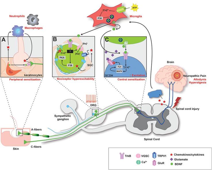
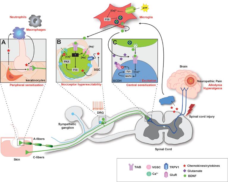
Pain is a sensory state resulting from complex integration of peripheral nociceptive inputs and central processing. Pain consists of adaptive pain that is acute and beneficial for healing and maladaptive pain that is often persistent and pathological. Pain is indeed heterogeneous, and can be expressed as nociceptive, inflammatory, or neuropathic in nature. Neuropathic pain is an example of maladaptive pain that occurs after spinal cord injury (SCI), which triggers a wide range of neural plasticity. The nociceptive processing that underlies pain hypersensitivity is well-studied in the spinal cord. However, recent investigations show maladaptive plasticity that leads to pain, including neuropathic pain after SCI, also exists at peripheral sites, such as the dorsal root ganglia (DRG), which contains the cell bodies of sensory neurons. This review discusses the important role DRGs play in nociceptive processing that underlies inflammatory and neuropathic pain. Specifically, it highlights nociceptor hyperexcitability as critical to increased pain states. Furthermore, it reviews prior literature on glutamate and glutamate receptors, voltage-gated sodium channels (VGSC), and brain-derived neurotrophic factor (BDNF) signaling in the DRG as important contributors to inflammatory and neuropathic pain. We previously reviewed BDNF's role as a bidirectional neuromodulator of spinal plasticity. Here, we shift focus to the periphery and discuss BDNF-TrkB expression on nociceptors, non-nociceptor sensory neurons, and non-neuronal cells in the periphery as a potential contributor to induction and persistence of pain after SCI. Overall, this review presents a comprehensive evaluation of large bodies of work that individually focus on pain, DRG, BDNF, and SCI, to understand their interaction in nociceptive processing.
Keywords: BDNF; Dorsal root ganglia; Nociceptors; Pain; Spinal cord injury; TrkB.
© 2024 The Authors.
Conflict of interest statement
The authors declare that they have no known competing financial interests or personal relationships that could have appeared to influence the work reported in this paper.
Figures
Similar articles
-
TrkB Agonist (7,8-DHF)-Induced Responses in Dorsal Root Ganglia Neurons Are Decreased after Spinal Cord Injury: Implication for Peripheral Pain Mechanisms.eNeuro. 2025 Jan 3;12(1):ENEURO.0219-24.2024. doi: 10.1523/ENEURO.0219-24.2024. Print 2025 Jan. eNeuro. 2025. PMID: 39753357 Free PMC article.
-
Role of the immune system in neuropathic pain.Scand J Pain. 2019 Dec 18;20(1):33-37. doi: 10.1515/sjpain-2019-0138. Scand J Pain. 2019. PMID: 31730538 Review.
-
Nociceptors as chronic drivers of pain and hyperreflexia after spinal cord injury: an adaptive-maladaptive hyperfunctional state hypothesis.Front Physiol. 2012 Aug 2;3:309. doi: 10.3389/fphys.2012.00309. eCollection 2012. Front Physiol. 2012. PMID: 22934060 Free PMC article.
-
Major Differences in Transcriptional Alterations in Dorsal Root Ganglia Between Spinal Cord Injury and Peripheral Neuropathic Pain Models.J Neurotrauma. 2023 May;40(9-10):883-900. doi: 10.1089/neu.2022.0238. Epub 2022 Oct 31. J Neurotrauma. 2023. PMID: 36178348 Free PMC article.
-
Regional Hyperexcitability and Chronic Neuropathic Pain Following Spinal Cord Injury.Cell Mol Neurobiol. 2020 Aug;40(6):861-878. doi: 10.1007/s10571-020-00785-7. Epub 2020 Jan 18. Cell Mol Neurobiol. 2020. PMID: 31955281 Free PMC article. Review.
Cited by
-
Decoding pain chronification: mechanisms of the acute-to-chronic transition.Front Mol Neurosci. 2025 Jun 26;18:1596367. doi: 10.3389/fnmol.2025.1596367. eCollection 2025. Front Mol Neurosci. 2025. PMID: 40642387 Free PMC article. Review.
-
Functional Role of Piezo1 in the Human Eosinophil Cell Line AML14.3D10: Implications for the Immune and Sensory Nervous Systems.Biomolecules. 2024 Sep 14;14(9):1157. doi: 10.3390/biom14091157. Biomolecules. 2024. PMID: 39334923 Free PMC article.
-
TrkB Agonist (7,8-DHF)-Induced Responses in Dorsal Root Ganglia Neurons Are Decreased after Spinal Cord Injury: Implication for Peripheral Pain Mechanisms.eNeuro. 2025 Jan 3;12(1):ENEURO.0219-24.2024. doi: 10.1523/ENEURO.0219-24.2024. Print 2025 Jan. eNeuro. 2025. PMID: 39753357 Free PMC article.
-
A perspective: neuraxial therapeutics in pain management: now and future.Front Pain Res (Lausanne). 2024 Dec 10;5:1505019. doi: 10.3389/fpain.2024.1505019. eCollection 2024. Front Pain Res (Lausanne). 2024. PMID: 39720319 Free PMC article.
-
Preclinical Animal Models to Investigate the Role of Nav1.7 Ion Channels in Pain.Life (Basel). 2025 Apr 12;15(4):640. doi: 10.3390/life15040640. Life (Basel). 2025. PMID: 40283194 Free PMC article. Review.
References
-
- Abrahamsson T., Chou C.Y.C., Li S.Y., Mancino A., Costa R.P., Brock J.A., Nuro E., Buchanan K.A., Elgar D., Blackman A.V., Tudor-Jones A., Oyrer J., Farmer W.T., Murai K.K., Sjöström P.J. Differential regulation of evoked and spontaneous release by presynaptic NMDA receptors. Neuron. 2017;96(4):839–855.e835. doi: 10.1016/j.neuron.2017.09.030. - DOI - PubMed
Publication types
Grants and funding
LinkOut - more resources
Full Text Sources
