

Three-Dimensional Heat Map: The OTA/AO Type 43C Pilon Fracture Lines Distribution
- PMID: 38314199
- PMCID: PMC10838051
- DOI: 10.2147/IJGM.S444977
Three-Dimensional Heat Map: The OTA/AO Type 43C Pilon Fracture Lines Distribution
Abstract
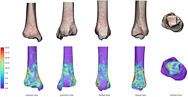
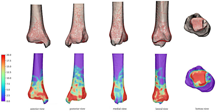
Background: This study aimed to create three-dimensional heat map and study the characteristic of fracture lines and represented fragments of OTA/AO type 43C pilon fractures.
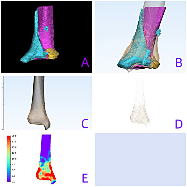
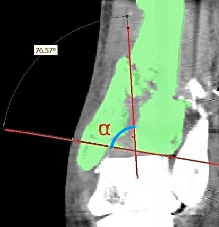
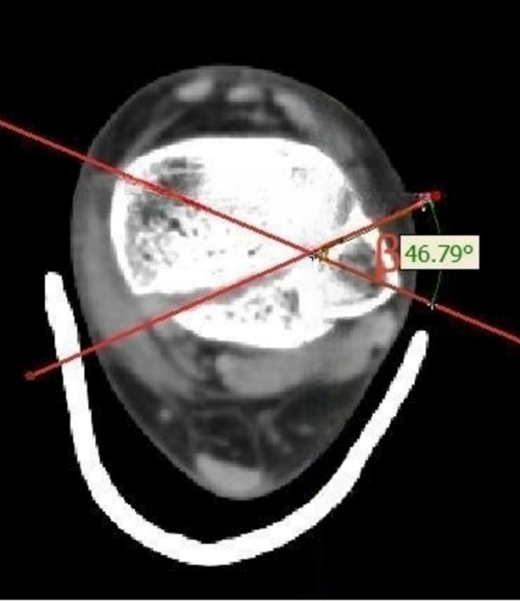
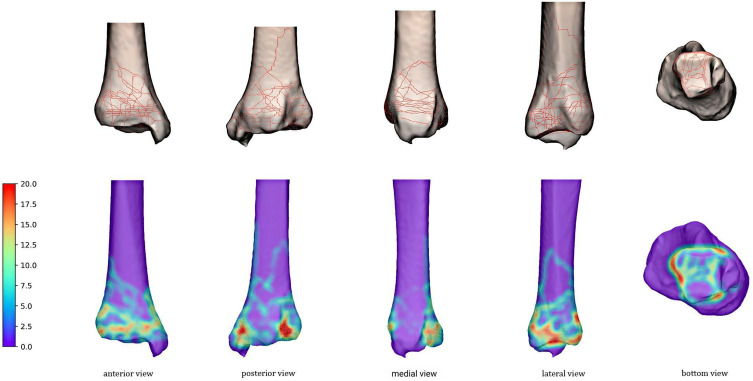
Methods: CT scan was performed in105 fractures diagnosed with OTA/AO type 43C pilon fractures between January 2017 and December 2022. Three-dimensional pilon fracture maps were created and converted into fracture heat maps. CT scan graphic parameters including the fracture line height, α angle, β angle, the ratio of the area and size of bone fragment represented by the fracture line to the total articular surface were measured.
Results: The study included 105 patients with 91 males and 14 females. The fractures included C1 (n=16), C2 (n=23), and C3 (n=66). There was no statistically different among the most parameters except in the fracture-line height of the anterior fracture line (p=0.03) and the sagittal fracture line (p=0.02) between C2 and C3 pilon fractures. The average size of the anterolateral fragment, occupied approximately 13.5% of the articular surface area, was (11.5±2.8) mm × (20.5±6.3) mm with the average height of 29.8 mm. The average size of the posterolateral fragment, occupied approximately 13.0% of the articular surface area, was (15.7±4.6) mm × (19.3±4.0) mm with the average height of 19.1 mm.
Conclusion: This study demonstrates that the articular surface fracture lines in the C type pilon fracture are formed by fixed main fracture lines. The understand of morphological and distribution characteristics of the fracture lines and size of fragments in OTA/AO type 43C pilon fractures would help the surgeons take suitable approach and fixation.
Keywords: classification; heat map; pilon fractures; three-dimensional reconstruction.
© 2024 Gao et al.
Conflict of interest statement
The author(s) report no conflicts of interest in this work.
Figures
Similar articles
-
Progress of fracture mapping technology based on CT three-dimensional reconstruction.Front Bioeng Biotechnol. 2024 Nov 6;12:1471470. doi: 10.3389/fbioe.2024.1471470. eCollection 2024. Front Bioeng Biotechnol. 2024. PMID: 39569162 Free PMC article. Review.
-
Ability of modern distal tibia plates to stabilize comminuted pilon fracture fragments: Is dual plate fixation necessary?Injury. 2016 Aug;47(8):1761-9. doi: 10.1016/j.injury.2016.05.026. Epub 2016 May 20. Injury. 2016. PMID: 27264277
-
Anterolateral distal tibia locking plate osteosynthesis and their ability to capture OTAC3 pilon fragments.Injury. 2018 Feb;49(2):409-413. doi: 10.1016/j.injury.2017.12.015. Epub 2017 Dec 16. Injury. 2018. PMID: 29305233
-
Fracture Line Morphology and a Novel Classification of Pilon Fractures.Orthop Surg. 2025 Feb;17(2):540-550. doi: 10.1111/os.14304. Epub 2024 Nov 23. Orthop Surg. 2025. PMID: 39579007 Free PMC article.
-
A Pilon Fracture With Fibular Head Dislocation Treated With the Use of 3D Preoperative Planning: A Case Report and Literature Review.J Foot Ankle Surg. 2021 Mar-Apr;60(2):404-407. doi: 10.1053/j.jfas.2020.09.014. Epub 2020 Oct 6. J Foot Ankle Surg. 2021. PMID: 33423890 Review.
Cited by
-
Progress of fracture mapping technology based on CT three-dimensional reconstruction.Front Bioeng Biotechnol. 2024 Nov 6;12:1471470. doi: 10.3389/fbioe.2024.1471470. eCollection 2024. Front Bioeng Biotechnol. 2024. PMID: 39569162 Free PMC article. Review.
References
LinkOut - more resources
Full Text Sources
Miscellaneous
