Case Reports
. 2024 Feb 1;16(2):e53422.
doi: 10.7759/cureus.53422. eCollection 2024 Feb.

Left Atrial Diverticula Present in the Right Lower Pulmonary Vein Thrombus Attachment Area

Affiliations
Case Reports

Left Atrial Diverticula Present in the Right Lower Pulmonary Vein Thrombus Attachment Area

Hidekazu Takeuchi. Cureus. .

Abstract

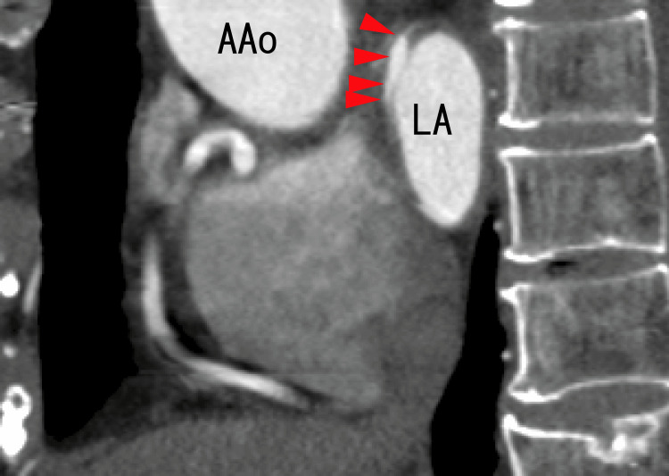
Left atrial diverticula (LADs) are thought to be associated with atrial fibrillation and an ischemic brain state. However, the mechanisms of LAD formation are unknown. Pulmonary vein thrombi (PVTs) can cause acute myocardial infarction (AMI) and ischemic stroke by releasing rather large particles. Additionally, PVTs can release much smaller particles, including neutrophil extracellular traps (NETs) and/or other components of NETs, such as DNA and histones. To treat these diseases, it may be crucial to know the specific traits of PVTs. However, these issues are not direct effects of PVTs on the left atrium (LA). It is unclear whether PVTs affect the LA directly. We checked the direct effects of PVTs on the LA using cardiac computed tomography (CT) and transesophageal echocardiography (TEE). The patient was a 73-year-old female with hypertension. TEE revealed extended LA thrombi from the right lower pulmonary vein, which were attached to the anterosuperior wall of the LA. Cardiac CT revealed the attaching area as a defect of enhancement and dimly revealed LAD with full thrombi on the attaching area. It was difficult to recognize the LAD at first; however, after one month of standard-dose heparin-warfarin treatment, the LAD was clearly detected using cardiac CT. LA thrombi could not be detected using cardiac CT.

Keywords: cardiac ct; left atrium diverticulum; pulmonary vein thrombosis; right lower pulmonary vein thrombi; tee.


Conflict of interest statement

The authors have declared that no competing interests exist.

Figures

Figure 1
Figure 1. TEE images demonstrating that LA thrombi extended from RLPV thrombi
TEE images demonstrating thrombi in the LA as thick line-like shapes connected to the entrance of the RUPV (arrows). The thrombi were not white and were thick and linear in shape. The blood flows from the RUPV are shown as mixtures of blue, yellow, and dark red areas. LA, left atrium; RLPV, right lower pulmonary vein; RUPV, right upper pulmonary vein
Figure 2
Figure 2. TEE images demonstrating thrombi in the left atrium
TEE images demonstrating thrombi in the LA as white line-like shapes that appeared to be connected to the entrance of the RUPV (arrows). The thrombi included a large white area and were linear in shape. The left end of the thrombi was attached to the wall of the LA (arrowheads). The blood flows from the RUPV and LUPV are shown as red areas. A portion of the blood flow from the RUPV is shown as blue areas. LA, left atrium; LUPV, left upper pulmonary vein; RLPV, right lower pulmonary vein; RUPV, right upper pulmonary vein
Figure 3
Figure 3. Axial images from an 80-MDCT scan revealing the RUPV, RLPV, and LA
There were no images of thrombi in the LA. There was a rather dark area in the RLPV, and there was a dark line in the center of the RUPV, indicating that there were thrombi in both the RUPV and the RLPV. Additionally, there was a strongly dark area in the LA (arrows), suggesting that both extended RLPV thrombi and the anterior wall of the LA were attached. LA, left atrium; RLPV, right lower pulmonary vein; RUPV, right upper pulmonary vein, MDCT, multidetector computed tomography
Figure 4
Figure 4. Axial images from an 80-MDCT scan revealing the RUPV, LLPV, and LA
There was a rather dark area (arrow), and there were some images of thrombi in the LA as the dark area. There were some enhanced areas on the anterior wall of the LA wall (arrowheads), suggesting that the diverticulum was full of thrombi. LA, left atrium; LAA, left atrial appendage; LLPV, left lower pulmonary vein; RUPV, right upper pulmonary vein; MDCT: multidetector computed tomography
Figure 5
Figure 5. Sagittal images from an 80-MDCT scan revealing the LA and LAD
There were more dark areas than LAs (arrowheads) on the anterior wall of the LA, suggesting that the diverticula were full of thrombi. The images of thrombi on the upper side of the LA are shown as darker areas. AAo, ascending aorta; LA, left atrium; MDCT, multidetector computed tomography
Figure 6
Figure 6. Axial images from an 80-MDCT scan obtained after one month of heparin and warfarin treatment
The location was similar to that of the 80-MDCT scan images in Figure 4. There was a diverticulum on the anterior wall of the LA (arrows). There was a little dark area in the LA, which was situated at the root of the diverticulum. The dark areas of the RUPV and LA became clearer, indicating that the thrombi had resolved. LA, left atrium; LAA, left atrial appendage; LLPV, left lower pulmonary vein; RUPV, right upper pulmonary vein; MDCT, multidetector computed tomography
Figure 7
Figure 7. Sagittal images from an 80-MDCT scan obtained after one month of heparin and warfarin treatment
The location was similar to that of the 80-MDCT scan images in Figure 5. There was a diverticulum on the anterior wall of the LA (arrowheads). The darker areas of the LA and LAD in Figure 5 became clearer, indicating that the thrombi had resolved. The distance between the anterior wall of the LAD and the anterior wall of the LA became shorter than that in Figure 5, indicating that the LAD decreased. AAo, ascending aorta; LA, left atrium; MDCT, multidetector computed tomography

References

    1. Are left atrial diverticula and left-sided septal pouches relevant additional findings in cardiac CT? Correlation between left atrial outpouching structures and ischemic brain alterations. Celik E, Pennig L, Laukamp KR, et al. Int J Cardiol. 2020;317:216–220. - PubMed
    1. Presence of accessory left atrial appendage/diverticula in a population with atrial fibrillation compared with those in sinus rhythm: a retrospective review. Troupis J, Crossett M, Scneider-Kolsky M, Nandurkar D. Int J Cardiovasc Imaging. 2012;28:375–380. - PubMed
    1. Cardiac CT assessment of left atrial accessory appendages and diverticula. Abbara S, Mundo-Sagardia JA, Hoffmann U, Cury RC. AJR Am J Roentgenol. 2009;193:807–812. - PubMed
    1. Left atrial diverticula in patients referred for radiofrequency ablation of atrial fibrillation: assessment of prevalence and morphologic characteristics by dual-source computed tomography. Peng LQ, Yu JQ, Yang ZG, et al. Circ Arrhythm Electrophysiol. 2012;5:345–350. - PubMed
    1. The incidence of left atrial diverticula in coronary CT angiography. Incedayi M, Öztürk E, Sonmez G, et al. Diagn Interv Radiol. 2012;18:542–546. - PubMed

Publication types

LinkOut - more resources