Case Reports
. 2024 Feb 3;16(2):e53526.
doi: 10.7759/cureus.53526. eCollection 2024 Feb.

Surgical Management of Congenital Pulmonary Airway Malformations (CPAM) in an Infant and a Toddler: Case Report Depicting Two Distinct Surgical Techniques With Successful Outcomes

Affiliations
Case Reports

Surgical Management of Congenital Pulmonary Airway Malformations (CPAM) in an Infant and a Toddler: Case Report Depicting Two Distinct Surgical Techniques With Successful Outcomes

Vishal V Bhende et al. Cureus. .

Abstract

Congenital pulmonary airway malformations (CPAM) compose the major part of congenital lung malformations (CLM) and have traditionally been treated by pulmonary lobectomy. In terms of surgical strategy, lobectomy has conventionally been the preferred treatment for CPAM localized to a single lobe. More recently, alternative approaches including lung-sparing resections (LSR), such as wedge or non-anatomic resections and segmentectomy, have been suggested. In asymptomatic CPAM early surgical resection is often shown to reduce infection and malignancy development. We describe two patients who were diagnosed with CPAM when being evaluated for respiratory tract infection. Patient 1 (P1) was a two-month-old infant weighing 4 kg with glucose-6-phosphate dehydrogenase (G6PD) deficiency and Patient 2 (P2) was a toddler aged one year, nine months weighing 9 kg. P1 underwent LSR for the CPAM diagnosed in the left upper lobe of the lung with conventional mechanical ventilation whilst right upper lobectomy was performed in P2 using one/single lung ventilation. In both cases, LSR and right upper lobectomy led to an uneventful postoperative recovery with no complications reported.

Keywords: congenital cystic adenomatoid malfomation; congenital cystic lung; congenital pulmonary airway malformation; conservative vs surgical management; infant; lung-sparing resection; segmental resection; surgical management; toddler; wedge resection.


Conflict of interest statement

The authors have declared that no competing interests exist.

Figures
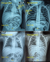
Figure 1
Figure 1. CXR pre-operative: (a) P1 showing CPAM cavity in left upper lobe of lung, (b) P2 showing CPAM cavity in right upper lobe of lung, (c) & (d) CXR postoperative
CXR - Chest X-ray; CPAM - Congenital pulmonary airway malformation; P - Patient Image Credit: Dr. Vishal V. Bhende
Figure 2
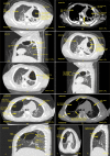
Figure 2. Case 1 (P1) (a) CECT scan of chest axial section in lung window (section 1) at the level of upper lobe showing a well-defined, thin-walled air containing cystic cavity lesion with surrounding lung parenchyma shows changes of passive collapse in left lung upper lobe. (b) CECT of section 1 in mediastinal window at level of upper lobe showing well-defined air-containing cavernous lesion with thin wall on CECT scan. (c) CECT of chest sagittal section in lung window showing air containing well-defined cystic lesion noted in left upper lobe with adjacent areas of collapse and left oblique fissure which separate left upper and lower lobe. (d) CECT of (section 1) in lung window at level of upper lobe showing a well-defined air-containing cavernous lesion in left upper lobe with upper lobar subsegmental bronchus appear in direct contact with cystic lesion and adjacent areas of passive collapse. (e) CECT of (section 1) showing well-defined air-containing cystic lesion in upper lobe of left lung measuring approximately 31 x 22 x 21 mm suggests CPAM (Type I) more likely. (f) CECT of chest sagittal section showing well-defined air-containing cystic lesion in upper lobe of left lung measuring approximately 31 x 22 x 21 mm suggests CPAM (Type I) more likely. Case 2 (P2) (g) HRCT scan of (section 1) at level of upper lobe showing a big, well-defined, oval, thin-walled, air-containing cystic cavity lesion in anterior segment of right upper lobe. There is thin oblique internal septation noted in antero-inferior portion of cavity. (h) HRCT scan of (section 1) in mediastinal window at level of upper lobe showing a big, well-defined, oval, thin walled, air containing cystic cavity lesion in anterior segment of right upper lobe. Apical segmental bronchus abuts medial wall of cystic lesion. (i) HRCT scan of chest sagittal section in lung window at right lung showing a big, well-defined, oval, thin-walled, air-containing cystic cavity lesion in anterior segment of right upper lobe. There is thin oblique internal septation noted in antero-inferior portion of cavity. The cystic lesion also abuts posteriorly to oblique fissure and inferiorly to horizontal fissure. (j & k) HRCT scan of chest axial and sagittal sections showing big, well-defined, oval, thin-walled, air-containing cystic lesion in anterior segment of right upper lobe measuring approximately 52.5 x 36.6 x 41.7 mm (AP X TX X SI) suggests congenital pulmonary airway malformation (Type I) more likely.
CECT - Contrast-enhanced computed tomography; HRCT - High-resolution computed tomography; CPAM - Congenital pulmonary airway malformation; AP - Antero-posterior; TX - Transverse; SI - Supero-inferior; P - Patient Image Credit: Dr. Dharmendra B. Parmar
Figure 3
Figure 3. Arndt Pediatric Endobronchial Blocker 5 Fr. (Cook Medical, Bloomington, IN, USA) with internal assembly (a & b)
Image Credit: Dr. Vishal V. Bhende
Figure 4
Figure 4. (a) Left limited thoracotomy exposure in Case 1 (b) Right postero-lateral thoracotomy exposure in Case 2
CPAM - Congenital pulmonary airway malformation; P - Patient Image Credit: Dr. Vishal V. Bhende
Figure 5
Figure 5. Surgical gross specimen of CPAM cavity from (a) left upper lobe of lung, (b) right upper lobe of lung, (inset-intact specimen) with cut-open specimen demonstrating cyst
CPAM - Congenital pulmonary airway malformation; CECT - Contrast enhanced computed tomography; AP - Antero-posterior; TX - Transverse; SI - Supero-inferior; P - Patient Image Credit: Dr. Vishal V. Bhende
Figure 6
Figure 6. Bronchial stapler provided by Endo-GIA (Covidien, Mansfield, MA, USA) used for closure of right upper lobe bronchus (a) Bronchial stapling gun (b) Cartridges
Image Credit: Dr. Vishal V. Bhende
Figure 7
Figure 7. Case 1 (a) Scanner view (4x magnification, H&E stain) showing fibro-collagenous cyst wall, lined by flattened epithelium (i), cyst cavity contains granulation tissue comprised of fibrous tissue, congested blood vessels, inflammatory cells (ii), (b) Low power view (10x magnification) shows adjacent alveolar tissue forming small, irregularly shaped airway spaces (i), lined by ciliated, cuboidal to columnar epithelium. The septa appear thickened and septal spaces appear dilated at places. Immature cartilage (ii) and bronchial glands (iii) are seen within the cyst wall, (c) High power view (40x magnification, H&E stain). Clusters of foamy macrophages and pneumocytes with vacuolated cytoplasm (arrows), Case 2 (d) Right side shows heavily congested lung parenchyma with collapsed alveoli (i), Left side shows adjacent lung tissue with mild congestion and focal alveolar enlargement (ii).
H&E - Hematoxylin and Eosin; P - Patient Image Credit: Dr. Zalak N. Parmar
Figure 8
Figure 8. Case 1 (a) HRCT Axial scan chest in mediastinal window, (b) Coronal HRCT scan chest in lung window showing areas of collapse with atelectasis noted in apico-posterior and anterior segments of left upper lobe with mild volume loss of left upper lobe with mild upward displacement of left oblique fissure. Case 2 (c) Postoperative axial HRCT scan of chest in mediastinal window showing mild fluid density areas with internal few small air foci noted in apical and anterior area adjacent to first and second intercostal space right side. There are few internal radiopaque surgical clips noted, (d) Postoperative coronal HRCT scan of chest in lung window showing stapler occluding right upper lobar bronchus. There are areas of adjacent sub-segmental collapse noted.
HRCT - High-resolution computed tomography; P - Patient Image Credit: Dr. Dharmendra B. Parmar
Figure 9
Figure 9. Categorisation of CPAM
CPAM - Congenital pulmonary airway malformation Image Credit: Dr. Vishal V. Bhende
Figure 10
Figure 10. Current prenatal treatments for CPAM
CPAM - Congenital pulmonary airway malformation; CVR - Cystic pulmonary airway malformation volume ratio; EXIT - Ex-utero intrapartum therapy Image Credit: Dr. Mathangi Krishnakumar
Figure 11
Figure 11. Algorithm of surgical indication for CPAM
CCAM - Congenital cystic adenoid malformation; CPAM - Congenital pulmonary airway malformation; EXIT - Ex-utero intrapartum therapy; VATS - Video-assisted thoracic surgery Image Credit: Dr. Vishal V. Bhende

References

    1. The natural history of prenatally diagnosed congenital cystic lung lesions: long-term follow-up of 119 cases. Cook J, Chitty LS, De Coppi P, Ashworth M, Wallis C. Arch Dis Child. 2017;102:798–803. - PubMed
    1. Congenital lung lesions. Zobel M, Gologorsky R, Lee H, Vu L. Semin Pediatr Surg. 2019;28:150821. - PubMed
    1. Congenital pulmonary airway malformation: a report of two cases. Bolde S, Pudale S, Pandit G, Ruikar K, Ingle SB. World J Clin Cases. 2015;3:470–473. - PMC - PubMed
    1. Congenital cystic adenomatoid malformation of the lung. Classification and morphologic spectrum. Stocker JT, Madewell JE, Drake RM. Hum Pathol. 1977;8:155–171. - PubMed
    1. Perinatally diagnosed asymptomatic congenital cystic adenomatoid malformation: to resect or not? Aziz D, Langer JC, Tuuha SE, Ryan G, Ein SH, Kim PC. J Pediatr Surg. 2004;39:329–334. - PubMed

Publication types

LinkOut - more resources