. 2024 Feb 13;121(7):e2314747121.
doi: 10.1073/pnas.2314747121. Epub 2024 Feb 5.

Precision treatment of viral pneumonia through macrophage-targeted lipid nanoparticle delivery

Affiliations

Precision treatment of viral pneumonia through macrophage-targeted lipid nanoparticle delivery

Gan Zhao et al. Proc Natl Acad Sci U S A. .

Abstract

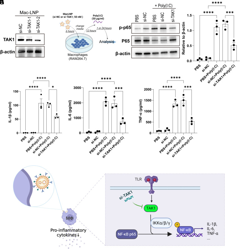
Macrophages are integral components of the innate immune system, playing a dual role in host defense during infection and pathophysiological states. Macrophages contribute to immune responses and aid in combatting various infections, yet their production of abundant proinflammatory cytokines can lead to uncontrolled inflammation and worsened tissue damage. Therefore, reducing macrophage-derived proinflammatory cytokine release represents a promising approach for treating various acute and chronic inflammatory disorders. However, limited macrophage-specific delivery vehicles have hindered the development of macrophage-targeted therapies. In this study, we screened a pool of 112 lipid nanoparticles (LNPs) to identify an optimal LNP formulation for efficient siRNA delivery. Subsequently, by conjugating the macrophage-specific antibody F4/80 to the LNP surface, we constructed MacLNP, an enhanced LNP formulation designed for targeted macrophage delivery. In both in vitro and in vivo experiments, MacLNP demonstrated a significant enhancement in targeting macrophages. Specifically, delivery of siRNA targeting TAK1, a critical kinase upstream of multiple inflammatory pathways, effectively suppressed the phosphorylation/activation of NF-kB. LNP-mediated inhibition of NF-kB, a key upstream regulator in the classic inflammatory signaling pathway, in the murine macrophage cell line RAW264.7 significantly reduced the release of proinflammatory cytokines after stimulation with the viral RNA mimic Poly(I:C). Finally, intranasal administration of MacLNP-encapsulated TAK1 siRNA markedly ameliorated lung injury induced by influenza infection. In conclusion, our findings validate the potential of targeted macrophage interventions in attenuating inflammatory responses, reinforcing the potential of LNP-mediated macrophage targeting to treat pulmonary inflammatory disorders.

Keywords: RNAi therapeutics; inhalation delivery; lipid nanoparticles; nanomedicine; pneumonia.


Conflict of interest statement

Competing interests statement:A.E.V., M.J.M., G.Z., and L.X. have filed a patent application based on this study. The other authors declare no competing interest.

Figures

Fig. 1.
Fig. 1.
Design of LNP formulations. (A) Study synopsis: LNP formulation design, screening, MacLNP assembly and validation, and in vivo application. (B) An antibody-modification strategy was utilized to conjugate antibody onto branched LNP surfaces for active targeting. Branched LNPs were formulated with branched lipidoids, phospholipids, cholesterol, PEG-lipids, and azide-PEG2K. The resulting branched LNPs were further incubated with DBCO-activated antibody for targeted LNPs formulation. (C) A list of seven amine cores and four branched tails was used for combinatorial design and synthesis of 28 branched lipidoids through a Michael-addition reaction.
Fig. 2.
Fig. 2.
In vitro screening of branched LNPs for siRNA delivery. (A) Branched LNPs were formulated through pipette mixing of an ethanol phase containing branched lipidoids (35%, molar ratio), phospholipids (DOPE or DSPC, 16%, molar ratio), cholesterol (46.5%, molar ratio), and PEG-lipids (C14PEG2K or DMG-PEG2K, 2.5%, molar ratio) and an aqueous phase containing siRNA. (B) A representative cryogenic transmission electron microscopy (cyro-TEM) image of branched LNP formulated by microfluidic mixing. (Scale bar: 100 nm.) (C) Hydrodynamic size of the representative branched LNP in B. (DG) Heat maps of GFP knockdown following treatment of HepG2-GFP cells with branched LNPs delivering siGFP at a concentration of 50 nM formulated under different parameters (n ≥ 3 replicates). (D) LNPs were formulated under parameter A with ethanol phase containing branched lipidoods, DOPE, cholesterol, and C14PEG2k. (E) LNPs were formulated under parameter B with ethanol phase containing branched lipidoids, DOPE, cholesterol, and DMG-PEG2K. (F) LNPs were formulated under parameter C with ethanol phase containing branched lipidoids, DSPC, cholesterol, and C14PEG2K. (G) LNPs were formulated under parameter D with ethanol phase containing branched lipidoids, DSPC, cholesterol, and DMG-PEG2K. (HJ) Structure–activity relationship (SAR) of branched LNPs for siRNA delivery. Relative hit rate (GFP silencing of >50%) was counted for GFP knockdown efficacy. (H) Relative hit rate of branched LNPs with different tertiary amine numbers per lipidoids. (I) Relative hit rate of branched LNPs with different branched degrees. (J) Relative hit rate of branched LNPs with different phospholipids and PEG-lipids. (K) Chemical structures of 4-O10b1 and 4-O10 lipidoids and their side view images generated from molecular dynamics simulations. (L) Critical packing parameters of 4-O10b1 and 4-O10 lipidoids calculated based on molecular dynamics simulations. (M) Hemolysis of LNPs with different dosages at pH 5.5 or 7.4. Red blood cells (RBCs) were incubated with LNPs at 37 °C for 1 h, and then, the supernatant was used to determine the adsorption at 540 nm. All data are presented as mean ± SEM (n = 3).
Fig. 3.
Fig. 3.
Validation of F4/80-conjugated LNP for macrophage delivery. (A) Schematic representation of the antibody-conjugated branched LNPs formulation. Azide LNP was formulated by microfluidics mixing of an ethanol phase containing 4-O10b1 lipidoid (35%, molar ratio), DOPE (16%, molar ratio), cholesterol (46.5%, molar ratio), C14PEG2K (2%, molar ratio), and azide-PEG (0.5%, molar ratio) with an aqueous phase containing siRNA. Subsequently, DBCO-activated monoclonal antibody (mAb) was incubated with the above LNP solution for clickable conjugation. (BD) (B) RAW264.7 cells were resuspended in 2 mL of Opi-MEM culture medium to obtain a single-cell suspension. These cells then mixed with LNP containing GFP mRNA (50 nM) as follows: LNP (no antibody conjugation), IgG LNP, and F4/80 LNP. The LNP-cell mixtures were then evenly distributed into a six-well plate. After 6 h, fluorescence microscopy was used to evaluate GFP expression. (C) Representative images of GFP expression from each group under the microscope. (Scale bar: 100 μm.) (D) Quantification of GFP cells, data are presented as means ± SEM (n = 3), calculated using one-way ANOVA, followed by Dunnett’s multiple comparison test, ***P < 0.001. (EG) (E) C57BL/6 mice were intranasally administered GFP-encapsulated LNPs, including LNP (without antibody conjugation), IgG LNP, and F4/80 LNP. Dexamethasone (DEX) was injected intraperitoneally (2 mg/kg; i.p.) 1 h prior to LNPs injection in all mice. Samples were collected 24 h later to observe GFP expression. (F) Representative images of macrophages (CD68+) expressing GFP from each group. (Scale bar: 100 μm.) (G) Quantification of GFP+ macrophages, data are presented as means ± SEM (n = 3), calculated using one-way ANOVA, followed by Dunnett’s multiple comparison test, **P < 0.01.
Fig. 4.
Fig. 4.
MacLNP delivery of TAK1 siRNA inhibits the release of proinflammatory cytokines in RAW264.7 cells. (A) RAW264.7 cells were transfected with siRNAs [50 nM, control siRNA (si-NC) and TAK1 siRNA (si-TAK1)] encapsulated in F4/80 LNP (MacLNP). TAK1 expression was analyzed using western blotting at 24 h posttransfection. (BG) (B) After transfection of RAW264.7 cells with MacLNP encapsulating siRNAs [50 nM; control siRNA (si-NC) and TAK1 siRNA (si-TAK1)], the cells were switched to complete culture medium after 6 h. Subsequently, they were cultured for an additional 12 h and then exposed to Poly(I:C) (50 μg/mL) for 1 h (for western blot analysis) or 24 h (supernatant collection for ELISA). (C) Western blot analysis was conducted to assess the phosphorylation levels of NF-κB p65. (D) Quantitative analysis was performed to measure the phosphorylation levels of NF-κB p65 relative to β-actin. ELISA was used to detect the levels of proinflammatory cytokines IL-1β (E), IL-6 (F), and TNF-α (G) in the supernatant. Data are presented as means ± SEM (n = 3), calculated using one-way ANOVA, followed by Dunnett’s multiple comparison test, ***P < 0.001, ****P < 0.0001. (H) The diagram shows how MacLNP delivers TAK1 (MAP3K) siRNA to reduce proinflammatory cytokine release from macrophages.
Fig. 5.
Fig. 5.
MacLNP delivery of TAK siRNA for the treatment of viral lung injury. (A) Timeline for MacLNP administration and sampling. C57BL/6 mice were treated with PBS or MacLNPs encapsulating control siRNA (si-NC) or TAK1 siRNA (si-TAK1) (1.25 mg/kg, intranasal) on days 15 and 20 postinfection, and samples were collected on day 25 postinfection. Dexamethasone-21-Phosphate (DEX) was injected intraperitoneally (i.p. 2 mg/kg) into the mice 1 h prior to PBS/LNP administration in all mice. (B and C) Total protein (B) and MPO activity (C) were quantified in BALF collected from mice which received treatment as described in A harvested on day 25 after infection. Data are represented as mean ± SEM (n = 4 to 5 mice/group), calculated using one-way ANOVA, followed by Dunnett’s multiple comparison test, *P < 0.05. (DF) The concentration of proinflammatory cytokines, IL-1β (D), IL-6 (E), and TNF-α (F) in BALF was measured by ELISA from mice which received treatment as described in A and harvested on day 25 after infection. Data are represented as mean ± SEM (n = 4 to 5 mice/group), calculated using one-way ANOVA, followed by Dunnett’s multiple comparison test, *P < 0.05. (G and H) (G) Upper: Tile scan images of H&E stain; Lower: clustered injury zone maps produced from upper H&E images. (Scale bars: 1 mm.) (H) Quantification of relative area in different injury zones in G. Data are represented as mean ± SEM (n = 4 to 5 mice/group), calculated using an unpaired two-tailed t test, *P < 0.05.

References

    1. Wynn T. A., Vannella K. M., Macrophages in tissue repair, regeneration, and fibrosis. Immunity 44, 450–462 (2016). - PMC - PubMed
    1. Watanabe S., Alexander M., Misharin A. V., Budinger G. R. S., The role of macrophages in the resolution of inflammation. J. Clin. Invest. 129, 2619–2628 (2019). - PMC - PubMed
    1. Chen W., et al. , Macrophage-targeted nanomedicine for the diagnosis and treatment of atherosclerosis. Nat. Rev. Cardiol. 19, 228–249 (2022). - PMC - PubMed
    1. McGonagle D., Sharif K., O’Regan A., Bridgewood C., The role of cytokines including interleukin-6 in COVID-19 induced Pneumonia and Macrophage Activation Syndrome-Like Disease. Autoimmun Rev. 19, 102537 (2020). - PMC - PubMed
    1. Suzuki T., et al. , Increased pulmonary GM-CSF causes alveolar macrophage accumulation mechanistic implications for desquamative interstitial pneumonitis. Am. J. Resp. Cell Mol. 62, 87–94 (2020). - PMC - PubMed

LinkOut - more resources