. 2024 Feb 13;121(7):e2320240121.
doi: 10.1073/pnas.2320240121. Epub 2024 Feb 5.

G-quadruplex DNA structure is a positive regulator of MYC transcription

Affiliations

G-quadruplex DNA structure is a positive regulator of MYC transcription

Isabel Esain-Garcia et al. Proc Natl Acad Sci U S A. .

Abstract

DNA structure can regulate genome function. Four-stranded DNA G-quadruplex (G4) structures have been implicated in transcriptional regulation; however, previous studies have not directly addressed the role of an individual G4 within its endogenous cellular context. Using CRISPR to genetically abrogate endogenous G4 structure folding, we directly interrogate the G4 found within the upstream regulatory region of the critical human MYC oncogene. G4 loss leads to suppression of MYC transcription from the P1 promoter that is mediated by the deposition of a de novo nucleosome alongside alterations in RNA polymerase recruitment. We also show that replacement of the endogenous MYC G4 with a different G4 structure from the KRAS oncogene restores G4 folding and MYC transcription. Moreover, we demonstrate that the MYC G4 structure itself, rather than its sequence, recruits transcription factors and histone modifiers. Overall, our work establishes that G4 structures are important features of transcriptional regulation that coordinate recruitment of key chromatin proteins and the transcriptional machinery through interactions with DNA secondary structure, rather than primary sequence.

Keywords: DNA; G-quadruplex; MYC; epigenetics; transcription.


Conflict of interest statement

Competing interests statement:S.B. is a founder, shareholder, and paid advisor and Director of Biomodal Ltd. and Inflex Ltd.; a founder, shareholder, and director of Elyx Ltd.; a founder and shareholder of RNAvate Ltd. L.M. is a paid consultant for Inflex Ltd. S.B. is a founder and shareholder (>5%) of Biomodal Ltd., Inflex Ltd., Elyx Ltd., and RNAvate Ltd.

Figures

Fig. 1.
Fig. 1.
Genome editing of the MYC G4. (A) Illustration of a G-tetrad formed from guanines by Hoogsteen base-pairing coordinated by a central cation (Left). Stacking of G-tetrads to form a G4 structure (Right). (B) Experimental overview of CRISPR editing of the MYC G4, indicating the core 27-bp sequence (Pu27) consisting of five G-runs and the extended 48-bp sequence, consisting of eight G-runs in total (Left). Schematic of the MYC upstream regulatory region. The MYC G4 is ~100 bp upstream of promoter P1 in the nuclease hypersensitive site (NHE III1). The wild-type MYC G4 and edited sequences are shown. MYC MUT and MUT MIN cell lines contain destabilizing mutations in the eight G-runs to abolish G4 folding. MUT CORE contains point mutations in the core five G-runs within the 27 bp sequence. For KRAS SWP cells, the MYC G4 is replaced with the KRAS G4, similar in structure but dissimilar in sequence. In MYC FLIP cells, the G4 is switched to the opposite (template) strand.
Fig. 2.
Fig. 2.
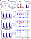
Structural perturbations of the MYC G4. (A) Circular dichroism (CD) spectra of MYC WT, MYC MUT, and KRAS SWP oligonucleotides (20 mM lithium cacodylate buffer, 10 mM KCl, pH 7.0). (B) CD melting curves for MYC and KRAS G4s, with Tm = 69 °C and Tm = 52 °C, respectively (20 mM lithium cacodylate buffer, 10 mM KCl, pH 7.0). (C) Kethoxal-assisted single-stranded-qPCR in MYC WT, MYC MUT, and KRAS SWP cells for the edited site, a positive G4-forming control (GAPDH) and G4-negative site (NANOG). Mean ± SD of MYC WT n = 1 and MYC MUT, KRAS SWP n = 3 independent biological samples measured twice as technical triplicates. (D) G4 CUT&Tag-qPCR with a G4 structure-specific antibody (BG4) for independently generated CRISPR clones for MYC WT, MYC MUT, and KRAS SWP cells. Mean ± SD of MYC MUT (N = 3), KRAS SWP (N = 2) independent clones, for n = 2 biological replicates, measured as technical triplicates. (E) BG4 CUT&Tag-qPCR (Left) for MYC WT, MYC MUT, and KRAS SWP cells relative to three G4s in control genes sites (RPA3, MAZ, RBBP4). G4 CUT&Tag sequencing profiles (Integrative Genomics Viewer, IGV tracks, Right) for the G4 edited site and the STAT3 control site. Mean ± SD, n = 3 independent biological samples. (F) as in (E) but with PDS Chem-map RT-qPCR (Left) and sequencing (Right). (G) as in (E) but with a nanobody raised against the MYC G4 (SG4). CUT&Tag-qPCR (Left) calculated against the G4-positive sites of RPA3, MAZ, and RBBP4. Sequencing tracks (Right) showing the SG4 and SG4 mutant CUT&Tag signals. SG4 signal is observed in MYC WT and KRAS SWP, while no binding is observed to MYC MUT or with SG4 mutant nanobody (see MYC WT, MYC MUT, and KRAS SWP). P-value: ns > 0.05, * ≤0.05, ** ≤0.01, *** ≤0.001, **** ≤0.0001.
Fig. 3.
Fig. 3.
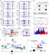
Loss of the MYC G4 structure abolishes MYC transcription from promoter P1. (A) Schematic of the G4 position in the upstream regulatory region of the MYC promoter P1. (B) RT-qPCR data for total MYC expression and P1 expression for multiple clones for each CRISPR-derived edit. Mean ± SD of MYC MUT (N = 3), KRAS SWP (N = 2) independent clones, for n = 2 biological replicates, measured as technical triplicates. (C) RNA-seq tracks (IGV) at the MYC P1 promoter site showing the absence of transcripts in MYC MUT cells compared to MYC WT and KRAS SWP cells. Blue bar indicates the P1 amplified region. (D) Volcano plots of RNA-seq data (Padj = 0.05, Log2 FC = 0.5). (E) Genomic binding profile (IGV track) showing BG4 CUT&Tag for the MYC FLIP cell line (Top). Gray regions indicate the location of G4 formation. RT-qPCR data for total MYC and P1 expression for the MYC FLIP genetic edit (Bottom). Mean ± SD, n = 3 independent biological samples. P-value: ns > 0.05, * ≤0.05, ** ≤0.01, *** ≤0.001, **** ≤0.0001.
Fig. 4.
Fig. 4.
The MYC G4 structure organizes the local chromatin landscape. (A) Genomic CUT&Tag binding profiles (IGV tracks) for SP1 and CNBP TFs (Top), MLL1 and 4 methyltransferases (Middle), and histone H3K4me1 and H3K4me3 methylation (Bottom) in MYC WT, MYC MUT, and KRAS SWP cells. (B) Affinity enrichment and western blot analysis for SP1 and CNBP proteins for double-strand (ds) and single-strand (ss) MYC G4, ss/ds MYC MUT, and ss/ds KRAS G4. (C) Genome-wide distribution of G4 CUT&Tag and MNase-seq data (Top). MNase-seq profile illustrating the deposition of a de novo nucleosome at the edited site (Bottom). (D) Genomic binding profiles (IGV tracks) showing RNAPII CUT&Tag for transcriptional initiation (RNAPIIS5P) and elongation (RNAPIIS2P), as well as nucleosome positioning (MNase-seq). (E) Suggested model where the G4 plays a central role to orchestrate a series of transcriptional events, including TF binding, molding the epigenetic landscape, organizing nucleosomes, and positioning RNAPII to regulate transcription in MYC.

References

    1. Varshney D., Spiegel J., Zyner K., Tannahill D., Balasubramanian S., The regulation and functions of DNA and RNA G-quadruplexes. Nat. Rev. Mol. Cell Biol. 21, 459–474 (2020). - PMC - PubMed
    1. Burge S., Parkinson G. N., Hazel P., Todd A. K., Neidle S., Quadruplex DNA: Sequence, topology and structure. Nucleic Acids Res. 34, 5402–5415 (2006). - PMC - PubMed
    1. Todd A. K., Johnston M., Neidle S., Highly prevalent putative quadruplex sequence motifs in human DNA. Nucleic Acids Res. 33, 2901–2907 (2005). - PMC - PubMed
    1. Huppert J. L., Balasubramanian S., G-quadruplexes in promoters throughout the human genome. Nucleic Acids Res. 35, 406–413 (2006). - PMC - PubMed
    1. Eddy J., Maizels N., Gene function correlates with potential for G4 DNA formation in the human genome. Nucleic Acids Res. 34, 3887–3896 (2006). - PMC - PubMed