

A late-life neurogenetic signature of exposure to combat stress - A monozygotic discordant twin study
- PMID: 38316103
- PMCID: PMC11113072
- DOI: 10.1016/j.jpsychires.2024.01.032
A late-life neurogenetic signature of exposure to combat stress - A monozygotic discordant twin study
Abstract
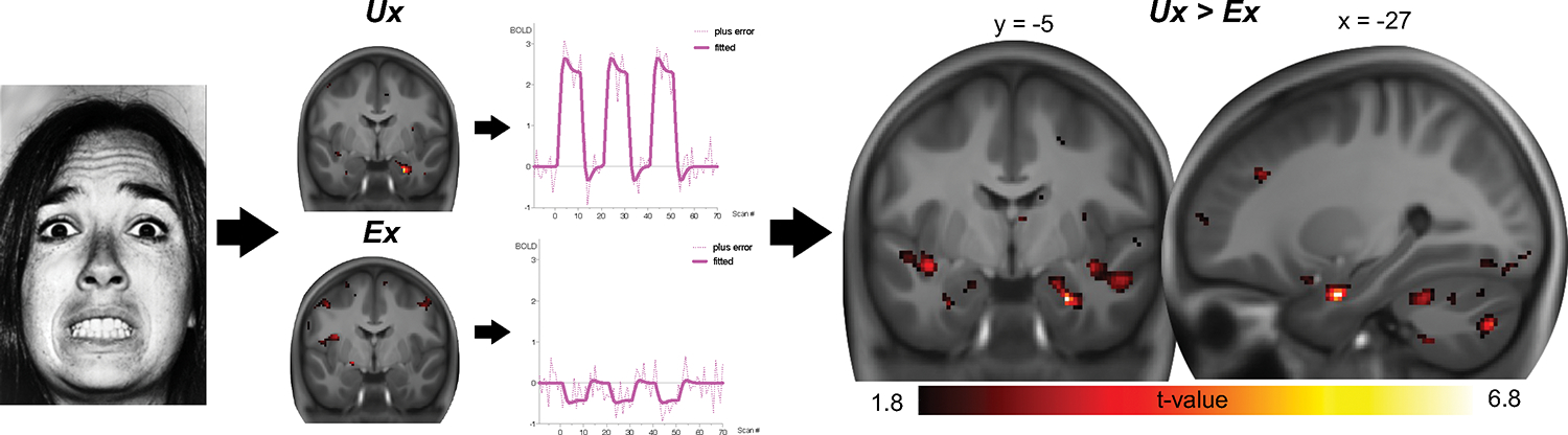
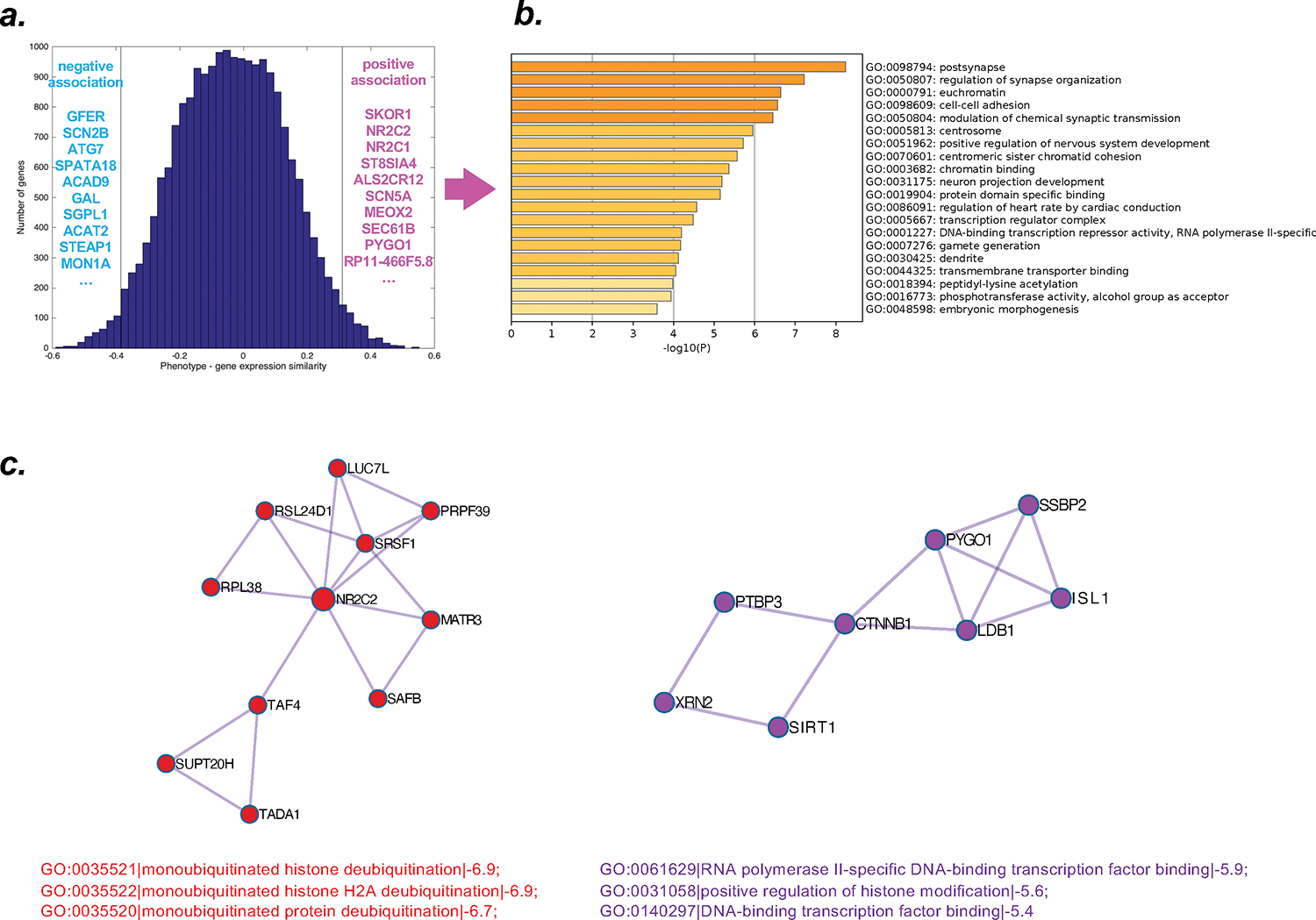
Animal models suggest that experiencing high-stress levels induces changes in amygdalar circuitry and gene expression. In humans, combat exposure has been shown to alter amygdalar responsivity and connectivity, but abnormalities have been indicated to normalize at least partially upon the termination of stress exposure. In contrast, other evidence suggests that combat exposure continues to exert influence on exposed individuals well beyond deployment and homecoming, as indicated by longitudinal psychosocial evidence from veterans, and observation of greater health decline in veterans late in life. Accordingly, the experience of combat stress early in life may affect amygdalar responsivity late in life, a possibility requiring careful consideration of the confounding effects of aging, genetic factors, and symptoms of post-traumatic stress disorder. Here, we investigated amygdalar responsivity in a unique sample of 16 male monozygotic (MZ) twin pairs in their sixties, where one but not the other sibling had been exposed to combat stress in early adulthood. Forty years after combat experience, a generally blunted amygdalar response was observed in combat-exposed veterans compared to their non-exposed twin siblings. Spatial associations between these phenotypical changes and patterns of gene expression in the brain were found for genes involved in the synaptic organization and chromatin structure. Protein-protein interactions among the set of identified genes pointed to histone modification mechanisms. We conclude that exposure to combat stress early in life continues to impact brain function beyond the termination of acute stress and appears to exert prolonged effects on amygdalar function later in life via neurogenetic mechanisms.
Keywords: Amygdala; Combat exposure; Stress; Trauma; Twins; fMRI.
Copyright © 2024. Published by Elsevier Ltd.
Conflict of interest statement
Declaration of Competing interest The authors declare no conflict of interest.
Figures
Similar articles
-
Amygdala volume in combat-exposed veterans with and without posttraumatic stress disorder: a cross-sectional study.Arch Gen Psychiatry. 2012 Oct;69(10):1080-6. doi: 10.1001/archgenpsychiatry.2012.73. Arch Gen Psychiatry. 2012. PMID: 23026958
-
Physiologic responses to sudden, loud tones in monozygotic twins discordant for combat exposure: association with posttraumatic stress disorder.Arch Gen Psychiatry. 2003 Mar;60(3):283-8. doi: 10.1001/archpsyc.60.3.283. Arch Gen Psychiatry. 2003. PMID: 12622661
-
Mixed-Handedness in Identical Twins Discordant for Combat Exposure in Vietnam: Relationship to Posttraumatic Stress Disorder.J Neuropsychiatry Clin Neurosci. 2016 Winter;28(1):45-8. doi: 10.1176/appi.neuropsych.15040090. Epub 2015 Sep 25. J Neuropsychiatry Clin Neurosci. 2016. PMID: 26404173
-
A twin study of the association of post-traumatic stress disorder and combat exposure with long-term socioeconomic status in Vietnam veterans.J Trauma Stress. 1995 Jan;8(1):111-24. doi: 10.1007/BF02105410. J Trauma Stress. 1995. PMID: 7712050
-
Identical but not the same: the value of discordant monozygotic twins in genetic research.Am J Med Genet B Neuropsychiatr Genet. 2010 Sep;153B(6):1134-49. doi: 10.1002/ajmg.b.31091. Am J Med Genet B Neuropsychiatr Genet. 2010. PMID: 20468073 Review.
References
-
- Breiter HC, Etcoff NL, Whalen PJ, Kennedy WA, Rauch SL, Buckner RL, Strauss MM, Hyman SE, Rosen BR, 1996. Response and habituation of the human amygdala during visual processing of facial expression. Neuron 17(5), 875–887. - PubMed
-
- Dannlowski U, Stuhrmann A, Beutelmann V, Zwanzger P, Lenzen T, Grotegerd D, Domschke K, Hohoff C, Ohrmann P, Bauer J, Lindner C, Postert C, Konrad C, Arolt V, Heindel W, Suslow T, Kugel H, 2012. Limbic scars: long-term consequences of childhood maltreatment revealed by functional and structural magnetic resonance imaging. Biol Psychiatry 71(4), 286–293. - PubMed
Publication types
MeSH terms
Grants and funding
LinkOut - more resources
Full Text Sources
Medical
