

Image annotation and curation in radiology: an overview for machine learning practitioners
- PMID: 38316659
- PMCID: PMC10844188
- DOI: 10.1186/s41747-023-00408-y
Image annotation and curation in radiology: an overview for machine learning practitioners
Abstract
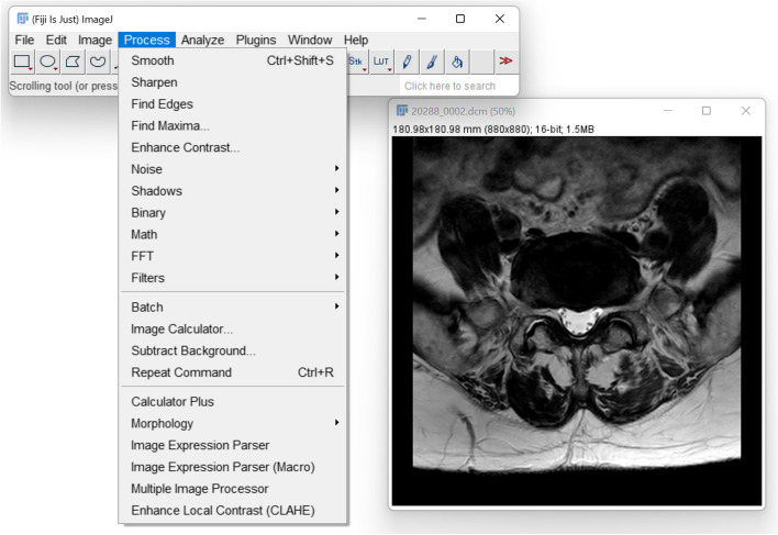
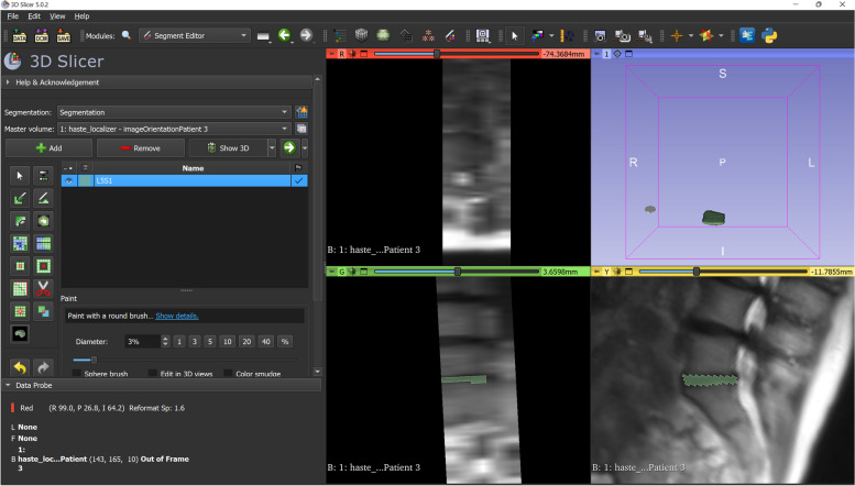
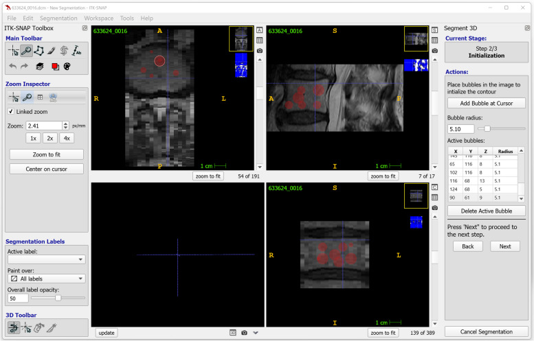
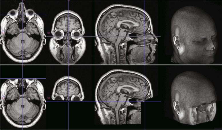
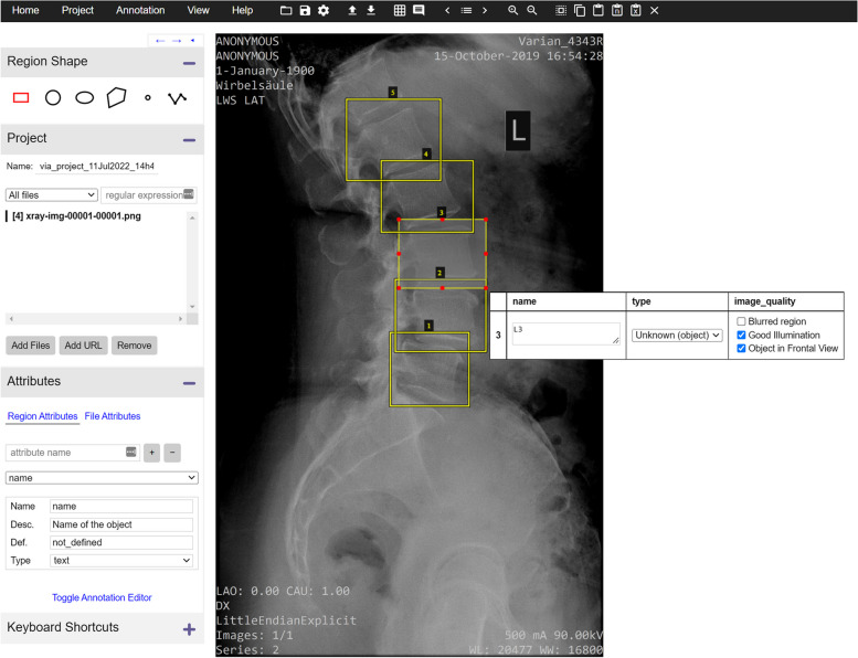
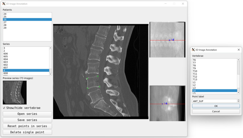
"Garbage in, garbage out" summarises well the importance of high-quality data in machine learning and artificial intelligence. All data used to train and validate models should indeed be consistent, standardised, traceable, correctly annotated, and de-identified, considering local regulations. This narrative review presents a summary of the techniques that are used to ensure that all these requirements are fulfilled, with special emphasis on radiological imaging and freely available software solutions that can be directly employed by the interested researcher. Topics discussed include key imaging concepts, such as image resolution and pixel depth; file formats for medical image data storage; free software solutions for medical image processing; anonymisation and pseudonymisation to protect patient privacy, including compliance with regulations such as the Regulation (EU) 2016/679 "General Data Protection Regulation" (GDPR) and the 1996 United States Act of Congress "Health Insurance Portability and Accountability Act" (HIPAA); methods to eliminate patient-identifying features within images, like facial structures; free and commercial tools for image annotation; and techniques for data harmonisation and normalisation.Relevance statement This review provides an overview of the methods and tools that can be used to ensure high-quality data for machine learning and artificial intelligence applications in radiology.Key points• High-quality datasets are essential for reliable artificial intelligence algorithms in medical imaging.• Software tools like ImageJ and 3D Slicer aid in processing medical images for AI research.• Anonymisation techniques protect patient privacy during dataset preparation.• Machine learning models can accelerate image annotation, enhancing efficiency and accuracy.• Data curation ensures dataset integrity, compliance, and quality for artificial intelligence development.
Keywords: Artificial intelligence; Data curation; Image processing (computer-assisted); Machine learning; Privacy.
© 2024. The Author(s).
Conflict of interest statement
F. Galbusera is a member of the European Radiology Experimental Editorial Board. He has not taken part in the review or selection process of this article.
The other author declares no competing interests.
Figures
Similar articles
-
A brief introduction to concepts and applications of artificial intelligence in dental imaging.Oral Radiol. 2021 Jan;37(1):153-160. doi: 10.1007/s11282-020-00468-5. Epub 2020 Aug 16. Oral Radiol. 2021. PMID: 32803680
-
Data Obfuscation Through Latent Space Projection for Privacy-Preserving AI Governance: Case Studies in Medical Diagnosis and Finance Fraud Detection.JMIRx Med. 2025 Mar 12;6:e70100. doi: 10.2196/70100. JMIRx Med. 2025. PMID: 40072927 Free PMC article.
-
De-Identification of Facial Features in Magnetic Resonance Images: Software Development Using Deep Learning Technology.J Med Internet Res. 2020 Dec 10;22(12):e22739. doi: 10.2196/22739. J Med Internet Res. 2020. PMID: 33208302 Free PMC article.
-
Artificial intelligence and machine learning for medical imaging: A technology review.Phys Med. 2021 Mar;83:242-256. doi: 10.1016/j.ejmp.2021.04.016. Epub 2021 May 9. Phys Med. 2021. PMID: 33979715 Free PMC article. Review.
-
Shallow and deep learning classifiers in medical image analysis.Eur Radiol Exp. 2024 Mar 5;8(1):26. doi: 10.1186/s41747-024-00428-2. Eur Radiol Exp. 2024. PMID: 38438821 Free PMC article. Review.
Cited by
-
Recent advances in deep learning for lymphoma segmentation: Clinical applications and challenges.Digit Health. 2025 Jul 28;11:20552076251362508. doi: 10.1177/20552076251362508. eCollection 2025 Jan-Dec. Digit Health. 2025. PMID: 40735544 Free PMC article. Review.
-
Current status and future direction of cancer research using artificial intelligence for clinical application.Cancer Sci. 2025 Feb;116(2):297-307. doi: 10.1111/cas.16395. Epub 2024 Nov 18. Cancer Sci. 2025. PMID: 39557634 Free PMC article. Review.
-
Foundation Models in Radiology: What, How, Why, and Why Not.Radiology. 2025 Feb;314(2):e240597. doi: 10.1148/radiol.240597. Radiology. 2025. PMID: 39903075 Review.
-
Mapping artificial intelligence models in emergency medicine: A scoping review on artificial intelligence performance in emergency care and education.Turk J Emerg Med. 2025 Apr 1;25(2):67-91. doi: 10.4103/tjem.tjem_45_25. eCollection 2025 Apr-Jun. Turk J Emerg Med. 2025. PMID: 40248473 Free PMC article. Review.
-
Developments in Deep Learning Artificial Neural Network Techniques for Medical Image Analysis and Interpretation.Diagnostics (Basel). 2025 Apr 23;15(9):1072. doi: 10.3390/diagnostics15091072. Diagnostics (Basel). 2025. PMID: 40361891 Free PMC article. Review.
References
Publication types
MeSH terms
LinkOut - more resources
Full Text Sources