

Anatomy-based correction of kidney PVE on [Formula: see text] SPECT images
- PMID: 38316677
- PMCID: PMC11266336
- DOI: 10.1186/s40658-024-00612-8
Anatomy-based correction of kidney PVE on [Formula: see text] SPECT images
Abstract
Background: In peptide receptor radionuclide therapy (PRRT), accurate quantification of kidney activity on post-treatment SPECT images paves the way for patient-specific treatment. Due to the limited spatial resolution of SPECT images, the partial volume effect (PVE) is a significant source of quantitative bias. In this study, we aimed to evaluate the performance and robustness of anatomy-based partial volume correction (PVC) algorithms to recover the accurate activity concentration of realistic kidney geometries on [Formula: see text]Lu SPECT images recorded under clinical conditions.
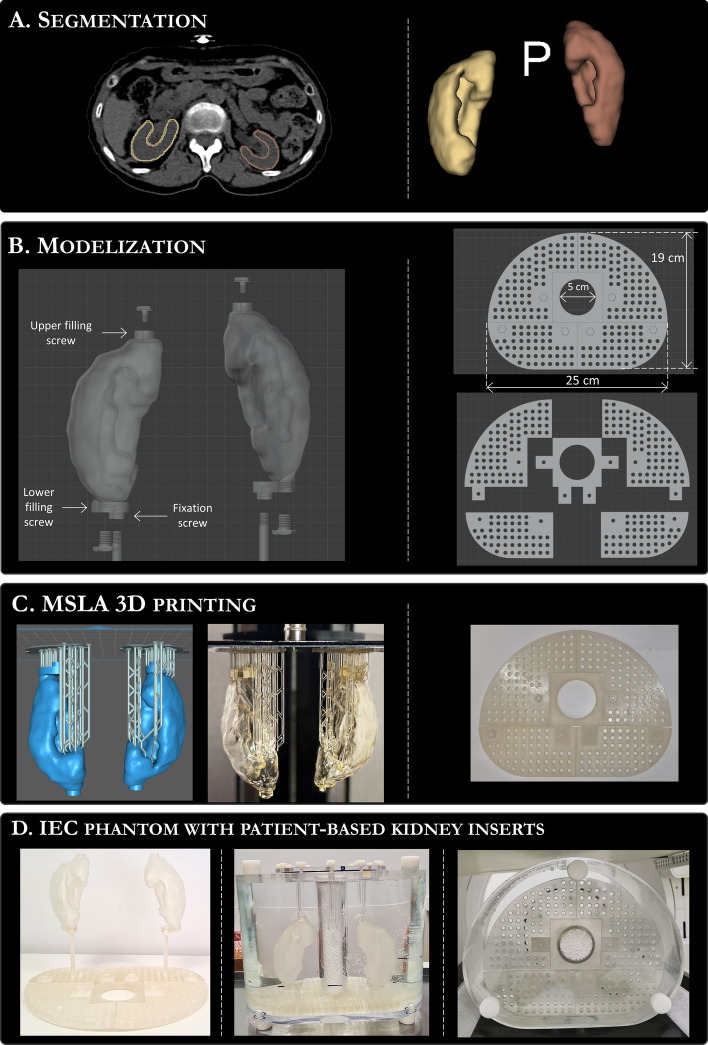
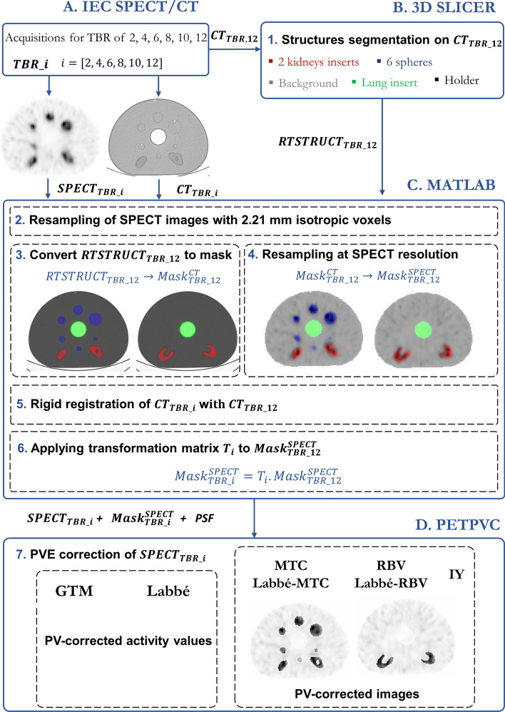
Methods: Based on the CT scan data from patients, three sets of fillable kidneys with surface-to-volume (S:V) ratios ranging from 1.5 to 2.8 cm-1, were 3D printed and attached in a IEC phantom. Quantitative [Formula: see text]Lu SPECT/CT acquisitions were performed on a GE Discovery NM CT 870 DR camera for the three modified IEC phantoms and for 6 different Target-To-Background ratios (TBRs: 2, 4, 6, 8, 10, 12). Two region-based (GTM and Labbé) and five voxel-based (GTM + MTC, Labbé + MTC, GTM + RBV, Labbé + RBV and IY) methods were evaluated with this data set. Additionally, the robustness of PVC methods to Point Spread Function (PSF) discrepancies, registration mismatches and background heterogeneity was evaluated.
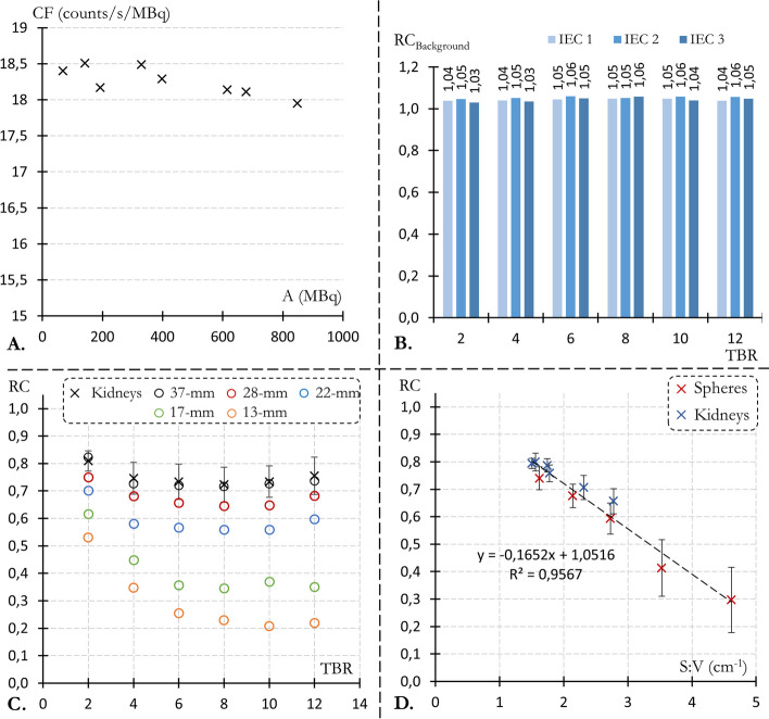
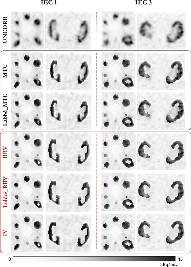
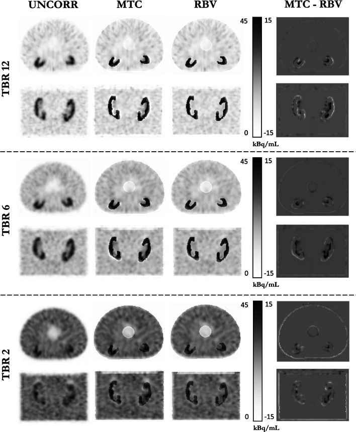
Results: Without PVC, the average kidney RCs across all TBRs ranged from 0.66 ± 0.05 (smallest kidney) to 0.80 ± 0.03 (largest kidney). For a TBR of 12, all anatomy-based method were able to recover the kidneys activity concentration with an error < 6%. All methods result in a comparable decline in RC restoration with decreasing TBR. The Labbé method was the most robust against PSF and registration mismatches but was also the most sensitive to background heterogeneity. Among the voxel-based methods, MTC images were less uniform than RBV and IY images at the outer edge of high uptake areas (kidneys and spheres).
Conclusion: Anatomy-based PVE correction allows for accurate SPECT quantification of the [Formula: see text]Lu activity concentration with realistic kidney geometries. Combined with recent progress in deep-learning algorithms for automatic anatomic segmentation of whole-body CT, these methods could be of particular interest for a fully automated OAR dosimetry pipeline with PVE correction.
Keywords: Lu; Partial volume correction; Partial volume effect; Peptide receptor radionuclide therapy; SPECT imaging.
© 2024. The Author(s).
Conflict of interest statement
The authors declare that they have no competing interests.
Figures


References
-
- Bodei L, Cremonesi M, Grana CM, Fazio N, Iodice S, Baio SM, Bartolomei M, Lombardo D, Ferrari ME, Sansovini M, Chinol M, Paganelli G. Peptide receptor radionuclide therapy with 177Lu-DOTATATE: the IEO phase I-II study. Eur J Nucl Med Mol Imaging. 2011;38:2125–35. 10.1007/s00259-011-1902-1. 10.1007/s00259-011-1902-1 - DOI - PubMed
-
- Zaknun JJ, Bodei L, Mueller-Brand J, Pavel ME, Baum RP, Hörsch D, O’Dorisio MS, O’Dorisiol TM, Howe JR, Cremonesi M, Kwekkeboom DJ. The joint IAEA, EANM, and SNMMI practical guidance on peptide receptor radionuclide therapy (PRRNT) in neuroendocrine tumours. Eur J Nucl Med Mol Imaging. 2013;40(5):800–16. 10.1007/s00259-012-2330-6. 10.1007/s00259-012-2330-6 - DOI - PMC - PubMed
-
- Danthala M, Kallur KG, Prashant GR, Rajkumar K, Raghavendra Rao M. 177Lu-DOTATATE therapy in patients with neuroendocrine tumours: 5 years’ experience from a tertiary cancer care centre in India. Eur J Nucl Med Mol Imaging. 2014;41(7):1319–26. 10.1007/s00259-014-2710-1. 10.1007/s00259-014-2710-1 - DOI - PubMed
-
- Strosberg J, El-Haddad G, Wolin E, Hendifar A, Yao J, Chasen B, Mittra E, Kunz PL, Kulke MH, Jacene H, Bushnell D, O’Dorisio TM, Baum RP, Kulkarni HR, Caplin M, Lebtahi R, Hobday T, Delpassand E, Van Cutsem E, Benson A, Srirajaskanthan R, Pavel M, Mora J, Berlin J, Grande E, Reed N, Seregni E, Öberg K, Lopera Sierra M, Santoro P, Thevenet T, Erion JL, Ruszniewski P, Kwekkeboom D, Krenning E. Phase 3 Trial of 177Lu-Dotatate for midgut neuroendocrine tumors. N Engl J Med. 2017;376(2):125–35. 10.1056/NEJMoa1607427. 10.1056/NEJMoa1607427 - DOI - PMC - PubMed
LinkOut - more resources
Full Text Sources
Miscellaneous
