. 2024 Feb 5;15(2):111.
doi: 10.1038/s41419-024-06475-2.

IL-17 promotes osteoclast-induced bone loss by regulating glutamine-dependent energy metabolism

Affiliations

IL-17 promotes osteoclast-induced bone loss by regulating glutamine-dependent energy metabolism

Renpeng Peng et al. Cell Death Dis. .

Abstract

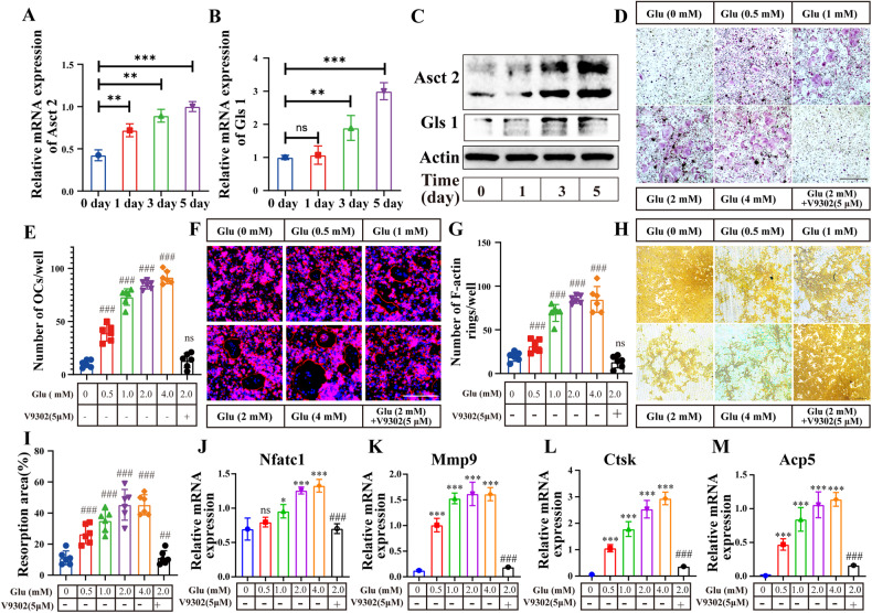
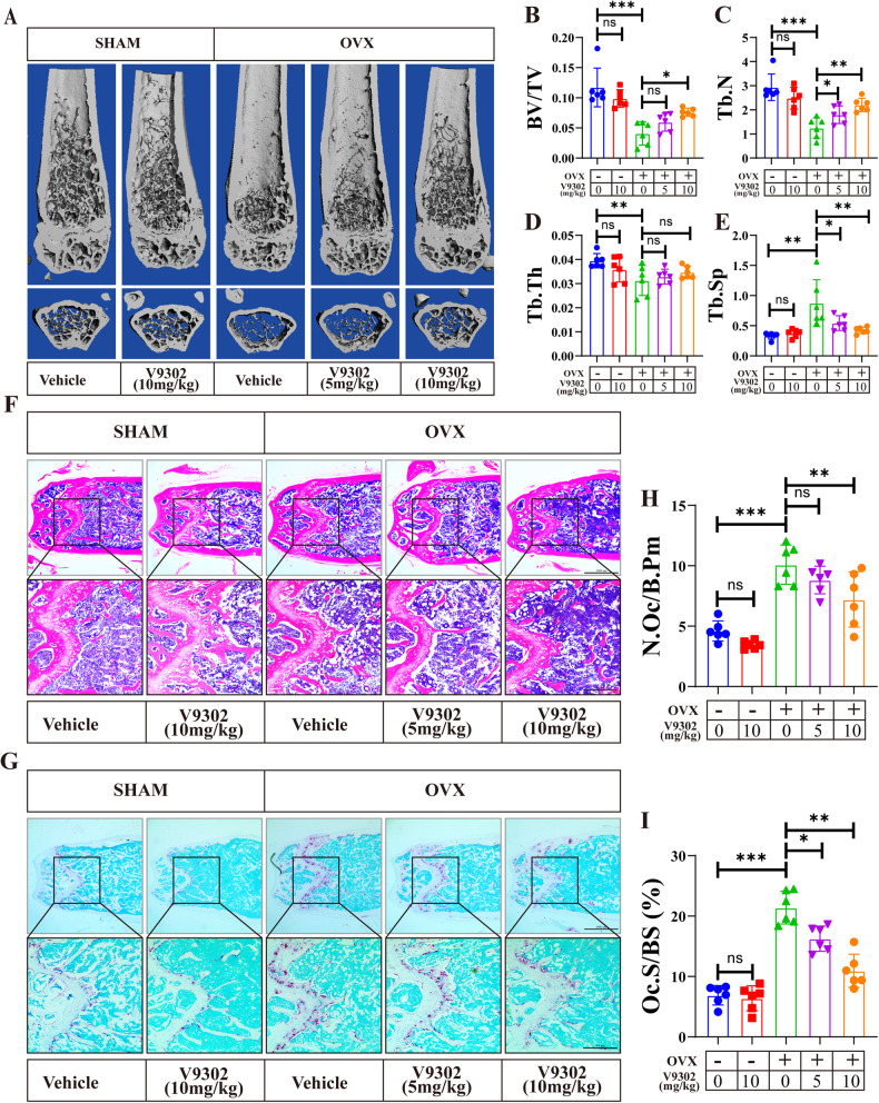
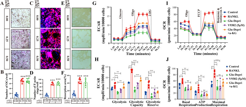
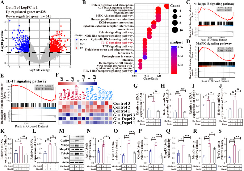
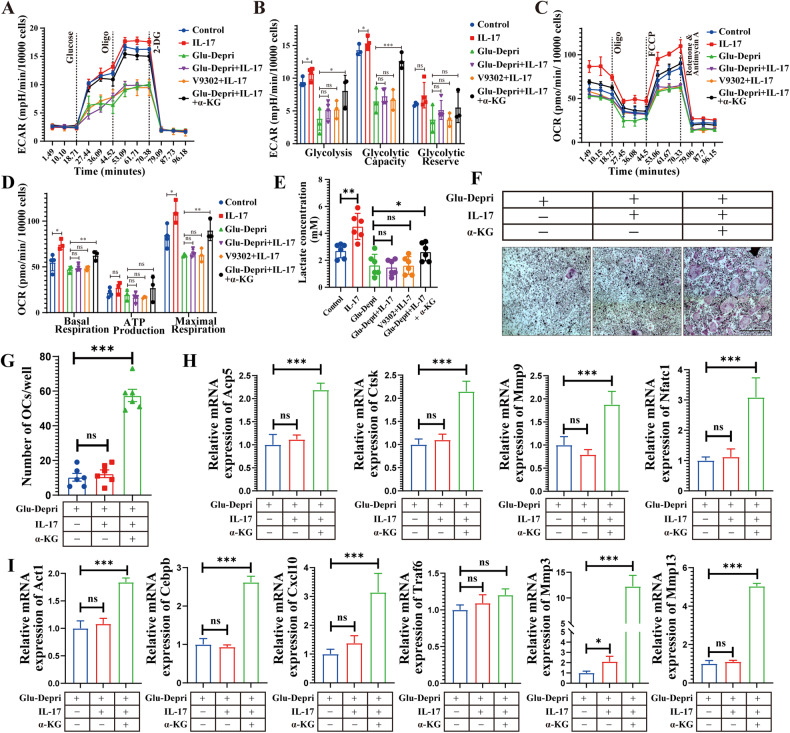
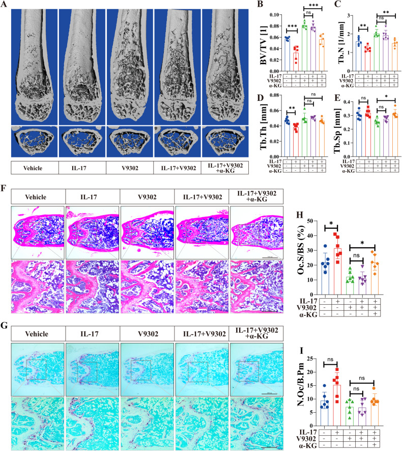
Osteoclasts consume an amount of adenosine triphosphate (ATP) to perform their bone resorption function in the development of osteoporosis. However, the mechanism underlying osteoclast energy metabolism has not been fully elucidated. In addition to glucose, glutamine (Glu) is another major energy carrier to produce ATP. However, the role of Glu metabolism in osteoclasts and the related molecular mechanisms has been poorly elucidated. Here we show that Glu is required for osteoclast differentiation and function, and that Glu deprivation or pharmacological inhibition of Glu transporter ASCT2 by V9302 suppresses osteoclast differentiation and their bone resorptive function. In vivo treatment with V9302 improved OVX-induced bone loss. Mechanistically, RNA-seq combined with in vitro and in vivo experiments suggested that Glu mediates the role of IL-17 in promoting osteoclast differentiation and in regulating energy metabolism. In vivo IL-17 treatment exacerbated OVX-induced bone loss, and this effect requires the participation of Glu or its downstream metabolite α-KG. Taken together, this study revealed a previously unappreciated regulation of IL-17 on energy metabolism, and this regulation is Glu-dependent. Targeting the IL-17-Glu-energy metabolism axis may be a potential therapeutic strategy for the treatment of osteoporosis and other IL-17 related diseases.


Conflict of interest statement

The authors declare no competing interests.

Figures

Fig. 1
Fig. 1. Glu is required for osteoclast differentiation.
AC The expression of Asct2 and Gls1 at the mRNA and protein level on day 1, 3, and 5 during OC differentiation. Statistical significance: *p < 0.05, **p < 0.01, ***p < 0.001; ns, no significance. D, E Glu regulate RANKL-induced osteoclastogenesis in a concentration-dependent manner. BMDMs were treated with different concentrations of Glu (0 mM,0.5 mM, 1 mM, 2 mM and 4 mM) or 5 μM V9302 for 5 days. TRAP-positive multinucleated (>3 nuclei) cells were counted as osteoclasts. *p < 0.05, **p < 0.01, ***p < 0.001. F, G BMDMs were seeded on 0.2% collagen-gel-coated 6-well plates and stimulated with 30 ng/ml M- CSF and 75 ng/ml RANKL for 6 days. Then, the cells were digested and seeded onto the Osteo Assay stripwell plates. Mature osteoclasts were treated with various concentrations of Glu or 5 μM V9302 for 5 days. F-actin staining was then performed. *p < 0.05, **p < 0.01, ***p < 0.001. H, I Pit formation assay in the Glu (0 mM,0.5 mM, 1 mM, 2 mM and 4 mM) or 5 μM V9302 treated group. Mature osteoclasts were cultured on Osteo Assay stripwell plates and treated with the medium containing RANKL and various concentrations of Glu for 2 days. The cells were then washed from the surface by using the 10% bleaching solution for 5 min. Resorption pits were captured with light microscopy and analyzed with Image J software. *p < 0.05, **p < 0.01, ***p < 0.001. JM BMDMs were cultured with the medium containing M- CSF, RANKL and various concentrations of Glu or were treated by 5 μM V9302 for 5 days. Relative mRNA expression levels of Nfatc1, Mmp9, Ctsk, and Acp5 versus β-Actin were quantified by qPCR. *compared to Glu (0 mM)/V9302 (0 μM) group; #compare to Glu (2 mM)/V9302(0 μM) group; *p < 0.05, **p < 0.01, ***p < 0.001. #p < 0.05, ##p < 0.01, ###p < 0.001.
Fig. 2
Fig. 2. Blocking Glu uptake by V9302 attenuates osteoclast-induced bone loss in OVX mice.
A Representative 3D-constructed images of the distal femurs of mice in each group. BE Quantitative analyses of bone structural parameters of the distal femurs, including bone volume/tissue volume (BV/TV), Tb.N (trabecular number), trabecular thickness (Tb.Th), and trabecular space (Tb.Sp). F Representative sections of the distal femurs were performed with H&E staining. G Representative sections of the distal femurs were performed with TRAP staining. H, I Quantitative analyses of histomorphometric bone parameters, including N.Oc/B.Pm and Oc.S/BS were performed, to reflect the formation of mature OCs on bone tissue slices. All data are presented as the mean ± SEM. *p < 0.05, **p < 0.01, ***p < 0.001. N = 6–8 per group.
Fig. 3
Fig. 3. Glu deficiency leads to impaired energy metabolism in osteoclasts, which can be rescured by α-ketoGlutarate (α-KG).
A, B BMDMs were cultured with the Glu deprived medium that containing M-CSF (30 ng/ml) and RANKL (75 ng/mL) for 5 days, as well as treated with indicated concentration of α-KG. TRAP-positive multinucleated (>3 nuclei) cells were counted as osteoclasts. C, D BMDMs were seeded on Osteo Assay stripwell plates and stimulated Glu deprived medium that containing M-CSF (30 ng/ml), RANKL (75 ng/mL) and indicated concentration of α-KG for 5 days. F-actin staining was then performed. E, F BMDMs were seeded on Osteo Assay stripwell plates and stimulated Glu deprived medium that containing M-CSF (30 ng/ml), RANKL (75 ng/mL) and indicated concentration of α-KG for 5 days. The cells were then washed from the surface by using the 10% bleaching solution for 5 min. Resorption pits were captured with light microscopy and analyzed with Image J software. G, H BMDMs seeded in Seahorse XF analyzer culture plates and treated as described in (A, B) Extracellular acidification rate (ECAR) were analyzed by XF Cell Mito Stress Assay. I, J BMDMs were seeded in Seahorse XF analyzer culture plates and treated as described in (A, B), Oxygen consumption rate (OCR) were analyzed by XF Cell Mito Stress Assay.
Fig. 4
Fig. 4. Glu is involved in the IL-17 signaling pathway during osteoclast differentiation.
A Volcano plot showing the differentially expressed genes between the control group and Glu deprivation group. Differentially expressed genes were identified by setting the threshold of |log2 (fold change)| to 1 and the P value to 0.05. B KEGG enrichment analysis showed the IL-17 signaling pathway were significantly altered after Glu deprivation. C, D GSEA analysis confirmed that Glu deprivation have no influence on MAPK and NFκB pathways. E GSEA analysis confirmed the inhibition of IL-17 signaling pathway by Glu deprivation. F Heatmap showing the downregulated genes of osteoclast markers and genes in the IL-17 signaling pathway. GS QPCR and Western blot assay examining the the expression of genes of osteoclast markers and genes in the IL-17 signaling pathway. *p < 0.05, **p < 0.01, ***p < 0.001; ns, no significance.
Fig. 5
Fig. 5. IL17 promotes osteoclast differentiation dependent on Glu.
BMDMs were cultured with the medium that containing M-CSF (30 ng/ml) and RANKL (75 ng/mL) for 5 days with or without Glu deprivation, as well as treated with IL-17 (0.1 ng/ml). A, B TRAP-positive multinucleated (>3 nuclei) cells were counted as osteoclasts. C, D F-actin staining was then performed. E, F BMDMs were seeded on Osteo Assay stripwell plates and cultured with the medium that containing M-CSF (30 ng/ml) and RANKL (75 ng/mL) for 5 days with or without Glu deprivation, as well as treated with IL-17(0.1 ng/ml). The cells were then washed from the surface by using the 10% bleaching solution for 5 min. Resorption pits were captured with light microscopy and analyzed with Image J software. G, H, I BMDMs were seeded in 6 well plates and treated as described. QPCR and Western blot assay examining the the expression of genes of osteoclast markers and genes in the IL-17 signaling pathway. *p < 0.05, **p < 0.01, ***p < 0.001; ns, no significance.
Fig. 6
Fig. 6. IL-17 regulates osteoclast energy metabolism via Glu.
BMDMs were cultured with the medium that containing M-CSF (30 ng/ml) and RANKL (75 ng/mL) for 5 days with or without Glu deprivation, as well as treated with indicated stimulation, including IL-17(0.1 ng/ml), V9302 (5 μM) and α-KG (0.5 mM). A, B BMDMs were seeded in Seahorse XF analyzer culture plates and treated as described. Extracellular acidification rate (ECAR) was analyzed by XF Cell Mito Stress Assay. C, D BMDMs were seeded in Seahorse XFp analyzer culture plates and treated as described. Oxygen consumption rate (OCR) were analyzed by XF Cell Mito Stress Assay. E BMDMs were seeded in 6 well plates and treated as described. The levels of lactate were analyzed through Lactate Assay kit. F, G BMDMs were cultured with the Glu deprived medium that containing M-CSF (30 ng/ml) and RANKL (75 ng/mL) for 5 days, as well as treated with or without IL-17 (0.1 ng/ml) and α-KG (0.5 mM). TRAP-positive multinucleated (>3 nuclei) cells were counted as osteoclasts. H, I QPCR and Western blot assay examining the the expression of gene of osteoclast marker and IL-17 signaling pathway. *p < 0.05, **p < 0.01, ***p < 0.001; ns, no significance.
Fig. 7
Fig. 7. The IL17-Glu axis increases bone loss in OVX mice.
A Representative 3D-constructed images of the distal femurs of mice in each group. BE Quantitative analyses of bone structural parameters of the distal femurs, including bone volume/tissue volume (BV/TV), Tb.N (trabecular number), trabecular thickness (Tb.Th), and trabecular space (Tb.Sp). F Representative sections of the distal femurs were performed with H&E staining. G Representative sections of the distal femurs were performed with TRAP staining. H, I Quantitative analyses of histomorphometric bone parameters, including N.Oc/B.Pm and Oc.S/BS were performed, to reflect the formation of mature OCs on bone tissue slices. All data are presented as the mean ± SEM. *p < 0.05, **p < 0.01, ***p < 0.001; ns, no significance. N = 6–8 per group.

References

    1. Dong Y, Kang H, Peng R, Song K, Guo Q, Guan H, et al. Global, regional, and national burden of low bone mineral density from 1990 to 2019: results from the global burden of disease study 2019. Front Endocrinol. 2022;13.870905. - PMC - PubMed
    1. Dong Y, Zhang Y, Song K, Kang H, Ye D, Li F. What was the epidemiology and global burden of disease of hip fractures from 1990 to 2019? results from and additional analysis of the global burden of disease study 2019. Clin Orthop Related Res. 2023;481:1209–20. doi: 10.1097/CORR.0000000000002465. - DOI - PMC - PubMed
    1. Dong Y, Peng R, Kang H, Song K, Guo Q, Zhao H, et al. Global incidence, prevalence, and disability of vertebral fractures: a systematic analysis of the global burden of disease study 2019. Spine J. 2022;22:857–68. doi: 10.1016/j.spinee.2021.12.007. - DOI - PubMed
    1. Kim JM, Lin C, Stavre Z, Greenblatt MB, Shim JH. Osteoblast-osteoclast communication and bone homeostasis. Cells. 2020;9:2073. doi: 10.3390/cells9092073. - DOI - PMC - PubMed
    1. Shen L, Hu G, Karner CM. Bioenergetic metabolism in osteoblast differentiation. Curr Osteoporos Rep. 2022;20:53–64. doi: 10.1007/s11914-022-00721-2. - DOI - PMC - PubMed

Publication types