. 2024 Feb 5;15(1):1076.
doi: 10.1038/s41467-024-44760-y.

Identification of myeloid-derived growth factor as a mechanically-induced, growth-promoting angiocrine signal for human hepatocytes

Affiliations

Identification of myeloid-derived growth factor as a mechanically-induced, growth-promoting angiocrine signal for human hepatocytes

Linda Große-Segerath et al. Nat Commun. .

Abstract

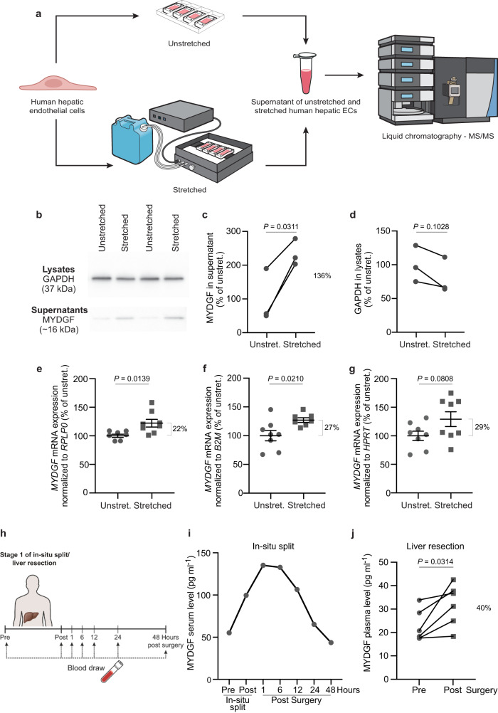
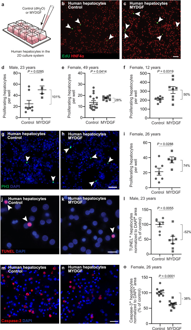
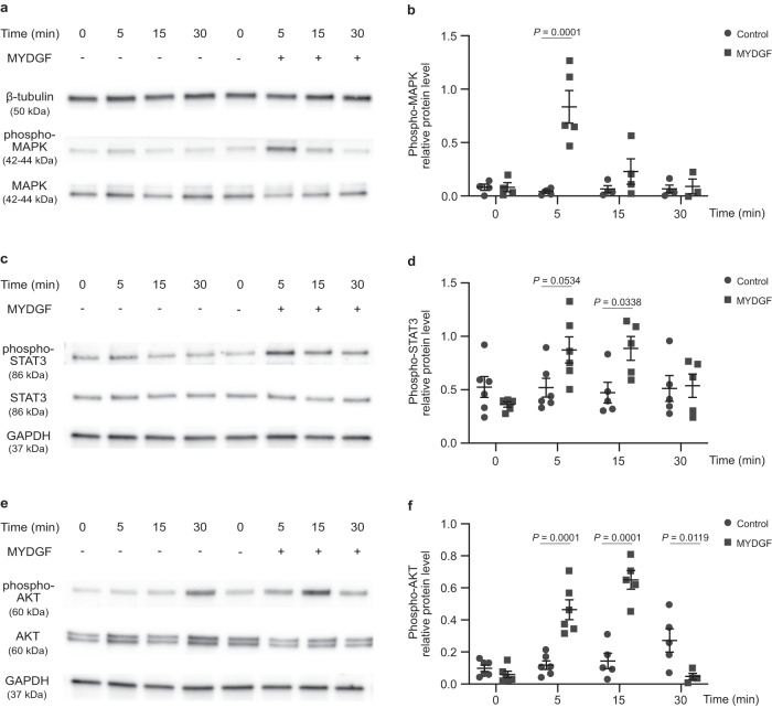
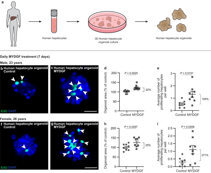
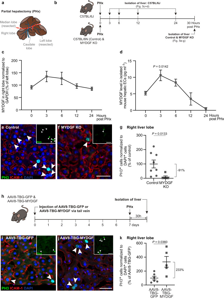
Recently, we have shown that after partial hepatectomy (PHx), an increased hepatic blood flow initiates liver growth in mice by vasodilation and mechanically-triggered release of angiocrine signals. Here, we use mass spectrometry to identify a mechanically-induced angiocrine signal in human hepatic endothelial cells, that is, myeloid-derived growth factor (MYDGF). We show that it induces proliferation and promotes survival of primary human hepatocytes derived from different donors in two-dimensional cell culture, via activation of mitogen-activated protein kinase (MAPK) and signal transducer and activator of transcription 3 (STAT3). MYDGF also enhances proliferation of human hepatocytes in three-dimensional organoids. In vivo, genetic deletion of MYDGF decreases hepatocyte proliferation in the regenerating mouse liver after PHx; conversely, adeno-associated viral delivery of MYDGF increases hepatocyte proliferation and MAPK signaling after PHx. We conclude that MYDGF represents a mechanically-induced angiocrine signal and that it triggers growth of, and provides protection to, primary mouse and human hepatocytes.


Conflict of interest statement

The authors declare no competing interests.

Figures

Fig. 1
Fig. 1. Identification of MYDGF as a mechanically-induced angiocrine signal.
a Experimental design for identification of angiocrine signals. Primary human hepatic endothelial cells (ECs) were mechanically stretched for 1.5 h (20% static stretch for 30 min plus 20% cyclic stretch (30 cycles/min) for 1 h), and the supernatants of unstretched and stretched hepatic ECs were analyzed by liquid chromatography tandem-mass spectrometry (LC-MS/MS). Myeloid-derived growth factor (MYDGF) was identified in the supernatants of mechanically stimulated hepatic ECs. b Representative Western blot images of supernatants (lower band) and lysates (upper band) of unstretched and stretched human hepatic ECs showing MYDGF (~16 kilodalton (kDa)) and GAPDH (37 kDa). Quantification of MYDGF levels in supernatants (c) and GAPDH levels in lysates (d) of unstretched and stretched hepatic ECs, shown as percentage of unstretched conditions; n = 3 independent experiments with 7 stretch chambers on average each. Two of the three independent experiments were performed with a male, 52-year-old human hepatic EC donor and one experiment with a 27-year-old female human hepatic EC donor. Quantification of MYDGF mRNA expression levels normalized to three housekeeping genes, i.e., RPLP0 (e), B2M (f) and HPRT (g) in lysates of unstretched and stretched hepatic ECs. n = 7 unstretched versus n = 8 stretched chambers (e), n = 8 unstretched versus n = 7 stretched chambers (f), n = 8 unstretched versus n = 8 stretched chambers (g). Primary human hepatic EC donor: male, 52 years. h Timeline of blood draw in a human patient pre and post in-situ split liver surgery. i MYDGF serum levels in a human patient at different timepoints (stage 1 of in situ-split/ liver resection); n = 1 patient (male, 70–80-year-old). j MYDGF plasma levels in six human patients pre and post liver resection (males, 54–89-year-old, body mass index (BMI) < 30 kg m-2). Data are presented as mean ± SEM (eg). P values were calculated using two-tailed paired Student’s t-test (c, d, j) and unpaired (eg) Student’s t-test with Welch’s correction. Source data are provided as a Source Data file.
Fig. 2
Fig. 2. MYDGF stimulates proliferation and prevents apoptosis of primary human hepatocytes in vitro.
a Schematic illustration of how human hepatocytes were treated with recombinant myeloid-derived growth factor (MYDGF). Representative laser scanning microscopy (LSM) images of human hepatocytes treated without (b) or with (c) recombinant MYDGF. White arrowheads point to proliferating cells stained for 5-ethynyl-2’-deoxyuridine (EdU, green); hepatocytes stained for hepatocyte nuclear factor 4α (HNF4α, red). Quantification of proliferating hepatocytes from three different human donors: (d) male, 23 years, n = 8 wells and n = 4 wells treated without or with MYDGF, respectively; (e) female, 49 years, n = 16 wells and n = 7 wells treated without or with MYDGF, respectively; (f) female, 12 years, n = 7 wells and n = 8 wells treated without or with MYDGF, respectively. Representative LSM images of human hepatocytes treated without (g) or with (h) recombinant MYDGF, stained for the proliferation marker phospho-Histone H3 (PH3, green). Cell nuclei were counterstained for DAPI (blue). i Quantification of proliferating human hepatocytes from a female, 26-year-old donor; n = 7 wells and n = 5 wells treated without or with MYDGF, respectively. Representative LSM images of human hepatocytes treated without (j) or with (k) recombinant MYDGF. Apoptotic cells were visualized by TUNEL (red) and cell nuclei were counterstained for DAPI (blue). l Quantification of TUNEL+ human hepatocytes from a male, 23-year-old donor; n = 7 wells each. Representative LSM images of human hepatocytes treated without (m) or with (n) recombinant MYDGF. Apoptotic cells were visualized by caspase-3 staining (red), and cell nuclei were counterstained for DAPI (blue). o Quantification of caspase-3+ human hepatocytes from a female, 26-year-old donor; n = 10 wells each. Scale bars: 100 µm (c), 50 µm (h, n), 20 µm (k). Data are presented as mean ± SEM. P values were calculated using two-tailed unpaired Student’s t-test with Welch’s correction. Source data are provided as a Source Data file.
Fig. 3
Fig. 3. MYDGF activates phosphorylation of MAPK, STAT3 and AKT.
Human hepatocytes were treated without or with recombinant myeloid-derived growth factor (MYDGF) for different times (0, 5, 15 and 30 min). a Western blots for phosphorylated mitogen-activated protein kinase (phospho-MAPK, 42–44 kDa; T202/Y204), MAPK (42–44 kDa) and β-tubulin (50 kDa) in lysates from human hepatocytes. b Quantification of phospho-MAPK protein levels normalized to MAPK and β-tubulin protein levels. n = 4 (0, 5, 15 and 30 min) control- versus n = 4 (0 and 15 min), n = 5 (5 min) and n = 3 (30 min) MYDGF-treated human hepatocytes. c Western blots for phosphorylated signal transducer and activator of transcription 3 (phospho-STAT3, 86 kDa; S727), STAT3 (86 kDa) and GAPDH (37 kDa). d Quantification of phospho-STAT3 protein levels normalized to STAT3 and GAPDH protein levels. n = 6 (0 and 5 min) and n = 5 (15 and 30 min) control- versus n = 5 (0, 15 and 30 min) and n = 6 (5 min) MYDGF-treated human hepatocytes. e Western blots for phospho-AKT (60 kDa; S473), AKT (60 kDa) and GAPDH (37 kDa). f Quantification of phospho-AKT protein levels normalized to AKT and GAPDH protein levels. n = 6 (0 and 5 min) and n = 5 (15 and 30 min) control- versus n = 6 (0 and 5 min), n = 5 (15 min) and n = 4 (30 min) MYDGF-treated human hepatocytes. Human hepatocyte donor: female, 26-year-old. Data are presented as mean ± SEM. P values were calculated using two-way ANOVA followed by Šidák´s post hoc test. Source data are provided as a Source Data file.
Fig. 4
Fig. 4. MYDGF enhances cell proliferation in human hepatocyte organoids.
a Schematic illustration of growing 3D organoids from human hepatocytes. Hepatocytes isolated from human liver were cultured in cell culture plates with low adhesion. After 3 days, organoids were formed, which could subsequently be treated with recombinant myeloid-derived growth factor (MYDGF). Representative laser scanning microscopy (LSM) images (maximum intensity projections) of human hepatocyte organoids treated daily without (b) or with (c) recombinant MYDGF. Proliferating cells were stained for 5-ethynyl-2’-deoxyuridine (EdU, green) and cell nuclei counterstained for DAPI (blue). Quantification of human organoid area (d) and proliferating human hepatocytes per well (e) in control- versus MYDGF-treated human hepatocyte organoids. n = 7 versus n = 8 wells with 5 organoids each. Representative LSM images (maximum intensity projections) of human hepatocyte organoids treated daily without (f) or with (g) recombinant MYDGF. Proliferating cells were stained for EdU (green) and cell nuclei counterstained for DAPI (blue). Quantification of human organoid area (h) and proliferating human hepatocytes per well (i) in control-versus MYDGF-treated human hepatocyte organoids. n = 8 wells with 5 organoids each (h) and n = 7 versus n = 8 wells with 5 organoids each (i). Donors: male, 23 years (be), and female, 26 years (fi). Scale bar: 50 µm (c, g). Data are presented as mean ± SEM. P values were calculated using two-tailed unpaired Student’s t-test with Welch’s correction. Source data are provided as a Source Data file.
Fig. 5
Fig. 5. MYDGF stimulates proliferation of hepatocytes in the mouse liver after partial hepatectomy.
a Anatomy of the mouse liver. b Schematic illustration of the experimental setup to determine myeloid-derived growth factor (MYDGF) expression kinetics and the effect of a global MYDGF knockout (KO) on hepatocyte proliferation after partial hepatectomy (PHx). c MYDGF protein levels in the right liver lobe relative to the respective left liver lobe of the same mouse, normalized to GAPDH protein levels; n = 5 (0 and 3 h), n = 4 (6 and 12 h) and n = 3 mice (24 h). d MYDGF protein levels in lysates of isolated mouse hepatic endothelial cells (ECs) from the right liver lobe; n = 5 (0 and 3 h), n = 4 (6 and 12 h) and n = 3 mice (24 h). Laser scanning microscopy (LSM) images of the right liver lobes of control (e) and MYDGF KO (f) mice. Phospho-Histone H3 (PH3, green, shown in inset), intercellular adhesion molecule-1 (ICAM-1, red), and DAPI (blue). g PH3+ (ICAM-1) cells normalized to DAPI+ cells in control versus MYDGF KO mice, shown as percentage of control. n = 8 control versus n = 5 MYDGF KO transversal sections of the right liver lobe. h An adeno-associated-virus of serotype 8 (AAV8) driving under the control of a thyroxine binding globulin (TBG) promoter, green fluorescent protein (GFP) or MYDGF, was injected into the tail vein of mice. Seven days post injection, a PHx was performed, and the livers were isolated another 30 h later. i, j LSM images of right mouse liver lobes. PH3 (green, shown in inset), ICAM-1 (red) and DAPI (blue). k PH3+ (ICAM-1) cells normalized to DAPI+ cells in AAV8-TBG-GFP versus AAV8-TBG-MYDGF mice, shown as percentage of AAV8-TBG-GFP. n = 6 AAV8-TBG-GFP versus n = 5 AAV8-TBG-MYDGF transversal sections of the right liver lobe. Scale bars: 50 µm (f, j). Data are presented as mean ± SEM. P values were calculated using one-way ANOVA followed by Dunnett’s post hoc test (c, d), and two-tailed unpaired Student’s t-test with Welch’s correction (g, k). Source data are provided as a Source Data file.

Similar articles

Cited by

References

    1. Michalopoulos GK, DeFrances MC. Liver regeneration. Science. 1997;276:60–66. doi: 10.1126/science.276.5309.60. - DOI - PubMed
    1. Fausto N. Liver regeneration. J. Hepatol. 2000;32:19–31. doi: 10.1016/S0168-8278(00)80412-2. - DOI - PubMed
    1. Taub R. Liver regeneration: from myth to mechanism. Nat. Rev. Mol. Cell Biol. 2004;5:836–847. doi: 10.1038/nrm1489. - DOI - PubMed
    1. Michalopoulos GK. Liver regeneration after partial hepatectomy: critical analysis of mechanistic dilemmas. Am. J. Pathol. 2010;176:2–13. doi: 10.2353/ajpath.2010.090675. - DOI - PMC - PubMed
    1. Lorenz L, et al. Mechanosensing by beta1 integrin induces angiocrine signals for liver growth and survival. Nature. 2018;562:128–132. doi: 10.1038/s41586-018-0522-3. - DOI - PubMed