

Deep brain stimulation of the central thalamus restores arousal and motivation in a zolpidem-responsive patient with akinetic mutism after severe brain injury
- PMID: 38316863
- PMCID: PMC10844373
- DOI: 10.1038/s41598-024-52267-1
Deep brain stimulation of the central thalamus restores arousal and motivation in a zolpidem-responsive patient with akinetic mutism after severe brain injury
Abstract
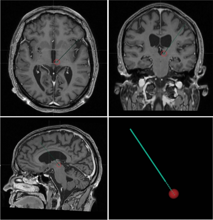
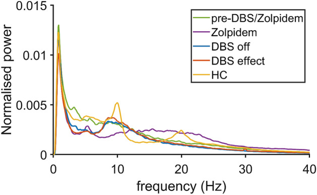
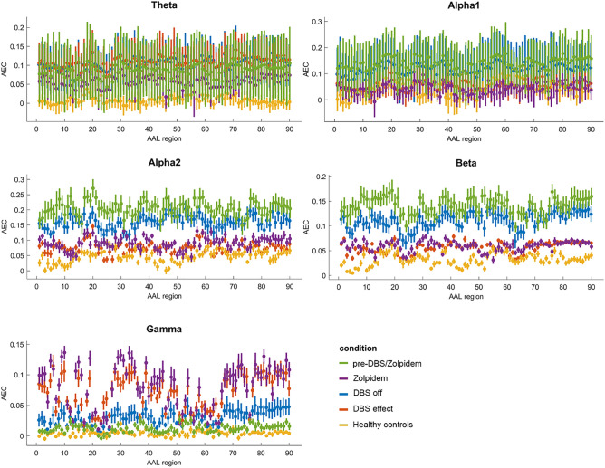
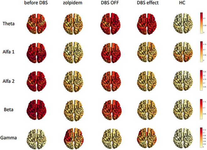
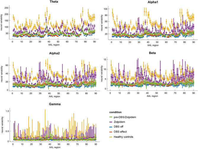
After severe brain injury, zolpidem is known to cause spectacular, often short-lived, restorations of brain functions in a small subgroup of patients. Previously, we showed that these zolpidem-induced neurological recoveries can be paralleled by significant changes in functional connectivity throughout the brain. Deep brain stimulation (DBS) is a neurosurgical intervention known to modulate functional connectivity in a wide variety of neurological disorders. In this study, we used DBS to restore arousal and motivation in a zolpidem-responsive patient with severe brain injury and a concomitant disorder of diminished motivation, more than 10 years after surviving hypoxic ischemia. We found that DBS of the central thalamus, targeted at the centromedian-parafascicular complex, immediately restored arousal and was able to transition the patient from a state of deep sleep to full wakefulness. Moreover, DBS was associated with temporary restoration of communication and ability to walk and eat in an otherwise wheelchair-bound and mute patient. With the use of magnetoencephalography (MEG), we revealed that DBS was generally associated with a marked decrease in aberrantly high levels of functional connectivity throughout the brain, mimicking the effects of zolpidem. These results imply that 'pathological hyperconnectivity' after severe brain injury can be associated with reduced arousal and behavioral performance and that DBS is able to modulate connectivity towards a 'healthier baseline' with lower synchronization, and, can restore functional brain networks long after severe brain injury. The presence of hyperconnectivity after brain injury may be a possible future marker for a patient's responsiveness for restorative interventions, such as DBS, and suggests that lower degrees of overall brain synchronization may be conducive to cognition and behavioral responsiveness.
© 2024. The Author(s).
Conflict of interest statement
The authors declare no competing interests.
Figures
Similar articles
-
Clinical and neurophysiological effects of central thalamic deep brain stimulation in the minimally conscious state after severe brain injury.Sci Rep. 2022 Jul 28;12(1):12932. doi: 10.1038/s41598-022-16470-2. Sci Rep. 2022. PMID: 35902627 Free PMC article.
-
Robust modulation of arousal regulation, performance, and frontostriatal activity through central thalamic deep brain stimulation in healthy nonhuman primates.J Neurophysiol. 2016 Nov 1;116(5):2383-2404. doi: 10.1152/jn.01129.2015. Epub 2016 Aug 31. J Neurophysiol. 2016. PMID: 27582298 Free PMC article.
-
Central thalamic deep brain stimulation to promote recovery from chronic posttraumatic minimally conscious state: challenges and opportunities.Neuromodulation. 2012 Jul;15(4):339-49. doi: 10.1111/j.1525-1403.2012.00458.x. Epub 2012 May 24. Neuromodulation. 2012. PMID: 22624587 Review.
-
Deep brain stimulation of the thalamus restores signatures of consciousness in a nonhuman primate model.Sci Adv. 2022 Mar 18;8(11):eabl5547. doi: 10.1126/sciadv.abl5547. Epub 2022 Mar 18. Sci Adv. 2022. PMID: 35302854 Free PMC article.
-
Moving toward a generalizable application of central thalamic deep brain stimulation for support of forebrain arousal regulation in the severely injured brain.Ann N Y Acad Sci. 2012 Aug;1265:56-68. doi: 10.1111/j.1749-6632.2012.06712.x. Epub 2012 Jul 26. Ann N Y Acad Sci. 2012. PMID: 22834729 Free PMC article. Review.
Cited by
-
Thalamic burst and tonic firing selectively indicate patients' consciousness level and recovery.Innovation (Camb). 2025 Feb 25;6(5):100846. doi: 10.1016/j.xinn.2025.100846. eCollection 2025 May 5. Innovation (Camb). 2025. PMID: 40432773 Free PMC article.
-
Shared subcortical arousal systems across sensory modalities during transient modulation of attention.Neuroimage. 2025 May 15;312:121224. doi: 10.1016/j.neuroimage.2025.121224. Epub 2025 Apr 16. Neuroimage. 2025. PMID: 40250641 Free PMC article.
-
A human brain network linked to restoration of consciousness after deep brain stimulation.Nat Commun. 2025 Jul 21;16(1):6721. doi: 10.1038/s41467-025-61988-4. Nat Commun. 2025. PMID: 40691170 Free PMC article.
-
Shared subcortical arousal systems across sensory modalities during transient modulation of attention.bioRxiv [Preprint]. 2024 Oct 1:2024.09.16.613316. doi: 10.1101/2024.09.16.613316. bioRxiv. 2024. Update in: Neuroimage. 2025 May 15;312:121224. doi: 10.1016/j.neuroimage.2025.121224. PMID: 39345640 Free PMC article. Updated. Preprint.
-
A human brain network linked to restoration of consciousness after deep brain stimulation.medRxiv [Preprint]. 2024 Oct 18:2024.10.17.24314458. doi: 10.1101/2024.10.17.24314458. medRxiv. 2024. Update in: Nat Commun. 2025 Jul 21;16(1):6721. doi: 10.1038/s41467-025-61988-4. PMID: 39484242 Free PMC article. Updated. Preprint.
References
-
- Bomalaski MN, Claflin ES, Townsend W, Peterson MD. Zolpidem for the treatment of neurologic disorders: A systematic review (vol 74, pg 1130, 2017) JAMA Neurol. 2017;74:1144–1144. - PubMed
MeSH terms
Substances
LinkOut - more resources
Full Text Sources
