. 2024 Apr;130(7):1187-1195.
doi: 10.1038/s41416-024-02583-0. Epub 2024 Feb 5.

Long-term activation of anti-tumor immunity in pancreatic cancer by a p53-expressing telomerase-specific oncolytic adenovirus

Affiliations

Long-term activation of anti-tumor immunity in pancreatic cancer by a p53-expressing telomerase-specific oncolytic adenovirus

Masashi Hashimoto et al. Br J Cancer. 2024 Apr.

Erratum in

Abstract

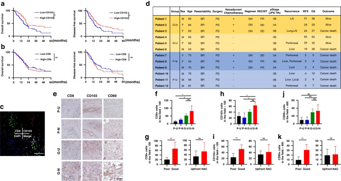
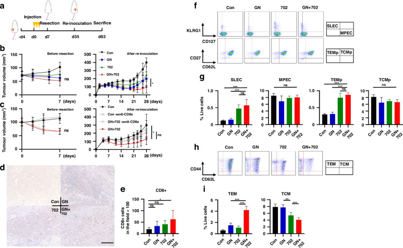
Background: Pancreatic cancer is an aggressive, immunologically "cold" tumor. Oncolytic virotherapy is a promising treatment to overcome this problem. We developed a telomerase-specific oncolytic adenovirus armed with p53 gene (OBP-702).

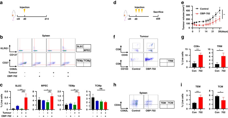
Methods: We investigated the efficacy of OBP-702 for pancreatic cancer, focusing on its long-term effects via long-lived memory CD8 + T cells including tissue-resident memory T cells (TRMs) and effector memory T cells (TEMs) differentiated from effector memory precursor cells (TEMps).

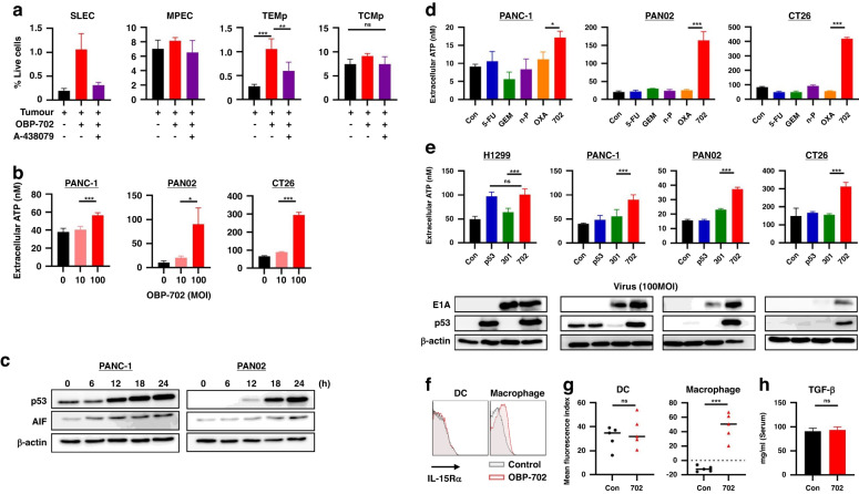
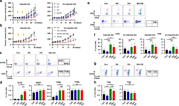
Results: First, in vitro, OBP-702 significantly induced adenosine triphosphate (ATP), which is important for memory T cell establishment. Next, in vivo, OBP-702 local treatment to murine pancreatic PAN02 tumors increased TEMps via ATP induction from tumors and IL-15Rα induction from macrophages, leading to TRM and TEM induction. Activation of these memory T cells by OBP-702 was also maintained in combination with gemcitabine+nab-paclitaxel (GN) in a PAN02 bilateral tumor model, and GN + OBP-702 showed significant anti-tumor effects and increased TRMs in OBP-702-uninjected tumors. Finally, in a neoadjuvant model, in which PAN02 cells were re-inoculated after resection of treated-PAN02 tumors, GN + OBP-702 provided long-term anti-tumor effects even after tumor resection.

Conclusion: OBP-702 can be a long-term immunostimulant with sustained anti-tumor effects on immunologically cold pancreatic cancer.


Conflict of interest statement

Yasuo Urata is President & CEO of Oncolys BioPharma, Inc., the manufacturer of OBP-702. Hiroshi Tazawa and Toshiyoshi Fujiwara are consultants of Oncolys BioPharma, Inc. The remaining authors declare no competing interests.

Figures

Fig. 1
Fig. 1. TCGA database analysis and CD8+ and CD103 + T cells in clinical specimens of pancreatic cancer.
a Kaplan–Meier curves for overall survival and disease-free survival for high-CD103 and low-CD103 pancreatic cancers from the TCGA database (n = 187). b Kaplan–Meier curves for overall survival and disease-free survival for high-CD8 and low-CD8 pancreatic cancers (n = 187). c Representative figures of immunofluorescent staining for CD8 (green), CD103 (red), DAPI (blue), and merge, for which the surgical specimen of patient 1 was used. Note that colocalization (yellow) of CD8 and CD103 means TRMs. Scale bar, 100 µm. d Clinicopathological characteristics of 12 patients with borderline resectable pancreatic ductal adenocarcinoma who underwent surgical resection are shown. G-N, G-U, P-N, and P-U mean good prognosis and NAC, good prognosis and upfront surgery, poor prognosis and NAC, and poor prognosis and upfront surgery, respectively. The cutoff period for a good or poor prognosis was defined as 24-month OS. e Twelve surgical specimens were subjected to immunohistochemical staining for CD8, CD103, and CD69. Representative images of each group (G-N, G-U, P-N, and P-U) are shown in (e). Scale bar, 200 µm. f, g The number of CD8+ TILs assessed in six different, randomly selected fields is compared among 4 groups (n = 3) (f), and between poor and good prognosis groups and between upfront and NAC groups (n = 6) (g). h, i The number of CD103+ TILs assessed in six different, randomly selected fields is compared among 4 groups (n = 3) (h), and between poor and good prognosis groups and between upfront and NAC groups (n = 6) (i). j, k The number of CD69+ TILs assessed in six different, randomly selected fields is compared among four groups (n = 3) (j), and between poor and good prognosis groups and between upfront and NAC groups (n = 6) (k). *P < 0.05, **P < 0.005, ***P < 0.001. RFS relapse-free survival, OS overall survival, BR borderline resectable, PD pancreatoduodenectomy, GN gemcitabine + nab-paclitaxel, PR partial response, SD stable disease, NAC neoadjuvant chemotherapy.
Fig. 2
Fig. 2. Activation of long-lived memory CD8 + T cells by OBP-702 in a PAN02 subcutaneous tumor model.
ac C57BL/6 mice bearing or not bearing PAN02 subcutaneous tumors were intratumorally treated with OBP-702 (1 × 108 PFU) or PBS once and sacrificed 14 days after treatment (a). Splenocytes collected from the harvested spleen were subjected to flow cytometry for SLECs, MPECs, TEMps, and TCMps. Representative figures of flow cytometry are shown in (b), in which CD3+/CD8+ cells are analyzed in the upper figures, and CD3+/CD8+/CD127+ cells are analyzed in the lower figures. Populations of SLECs, MPECs, TEMps, and TCMps are compared among control, OBP-702 vaccination, tumor inoculation, and OBP-702 treatment to tumor (n = 3–4) (c). d, e C57BL/6 mice bearing PAN02 subcutaneous tumors were intratumorally treated with PBS or OBP-702 (5.0 × 107 PFU) three times in a week and sacrificed 28 days after the initial treatment (d). Tumor volume was monitored after treatment by OBP-702 or PBS (n = 5) (e). f, g PAN02 tumors harvested at 28 days after the initial treatment with OBP-702 were subjected to flow cytometry for CD8+ cells and TRMs. Representative figures of flow cytometry are shown in (f), in which live cells are analyzed in the upper figures, and CD8+/CD45+ cells are analyzed in the lower figures. Populations of CD8+ cells and TRMs are compared between control and OBP-702 (n = 3–5) (g). h, i The spleens harvested 28 days after the initial treatment with OBP-702 were subjected to flow cytometry for TEMs and TCMs. Representative figures of flow cytometry are shown in (h), in which CD3+/CD8+ cells are analyzed. Populations of TEMs and TCMs are compared between control and OBP-702 (n = 5) (i). *P < 0.05, **P < 0.005, ***P < 0.001. SLEC short-lived effector T cell, MPEC memory precursor effector cell, TEMp effector memory precursor cell, TCMp central memory precursor cell, TRM tissue-resident memory T cell, TEM effector memory T cell, TCM central memory T cell, Con control, 702 OBP-702.
Fig. 3
Fig. 3. Stimulation of long-term immunity by OBP-702 via virus infection and p53 production.
a A-438079 was intraperitoneally injected into mice 6 h before OBP-702 treatment to PAN02 tumors that had been treated for 6 days to block P2RX7 receptors, and populations of SLECs, MPECs, TEMps, and TCMps in the splenocytes were analyzed by flow cytometry 7 days after treatment (n = 3–5). b Extracellular ATP secreted from PANC-1, PAN02, and CT26 cells treated with OBP-702 (0, 10, and 100 MOI) was measured with a luminescence assay 24 h after treatment (n = 3–5). c Whole cell lysates of PANC-1 and PAN02 cells collected at the indicated time points (0, 6, 12, 18, 24 h) after OBP-702 treatment (100 MOI) were subjected to western blotting for p53, AIF, and β-actin. d Extracellular ATP secreted from PANC-1, PAN02, and CT26 cells after monotherapy with chemotherapeutic agents (5-FU, GEM, nab-PTX, and OXA) and OBP-702 at the IC50 dose was measured with a luminescence assay 24 h after treatment (n = 3). e Extracellular ATP secreted from H1299, PANC-1, PAN02, and CT26 cells after treatment with Ad-p53, OBP-301, or OBP-702 at 100 MOI was measured with a luminescence assay 24 h after treatment (n = 3–5). Western blots of p53 and E1A are shown corresponding to the upper graph. f, g The spleens harvested 9 days after the initial treatment with OBP-702 were subjected to flow cytometry for IL-15-Rα on the surface of DCs (CD11b+/CD11c+/MHC-Class II+) and macrophages (CD11b+ except DC). Representative figures of flow cytometry are shown in (f), and the mean expression levels of IL-15-Rα are compared between control and OBP-702 (n = 5) (g). h The ELISA data show the murine serum TGF-β1 levels collected from the mice treated with PBS or OBP-702 7 days after treatment (n = 5). *P < 0.05, **P < 0.005, ***P < 0.001. SLEC short-lived effector T cell, MPEC memory precursor effector cell, TEMp effector memory precursor cell, TCMp central memory precursor cell, ATP adenosine triphosphate, MOI multiplicity of infection, AIF apoptosis-inducing factor, Con control, 5-FU 5-fluorouracil, GEM gemcitabine, n-P nab-paclitaxel, OXA oxaliplatin, 702 OBP-702, 301 OBP-301, DC dendritic cell.
Fig. 4
Fig. 4. Abscopal effect of combination therapy of GEM and nab-PTX with OBP-702 in a PAN02 bilateral subcutaneous tumor model.
a C57BL/6 mice bilaterally bearing PAN02 subcutaneous tumors were treated with GN (GEM 50 mg/kg, nab-PTX 5 mg/kg) intraperitoneally and/or OBP-702 (5 × 107 PFU) intratumorally once a week for 3 weeks, and sacrificed 34 days after initial treatment. OBP-702 was injected into the bigger tumor. Tumor volume was monitored (n = 7). The left figure shows tumor volume of the OBP-702-injected side, and the right figure shows the tumor volume of the OBP-702-uninjected side. b Anti-CD8α antibody was additionally injected into the peritoneal cavity once a week for 4 weeks, and tumor volume of control and OBP-702 with or without anti-CD8α antibody was monitored until 28 days after the initial treatment (n = 3–7). c, d The spleens harvested 14 days after the initial treatment were subjected to flow cytometry for SLECs, MPECs, TEMps, and TCMps. Representative figures of flow cytometry are shown in (c), in which CD3+/CD8+ cells are analyzed in the upper figures, and CD3+/CD8+/CD127+ cells are analyzed in the lower figures. Populations of SLECs, MPECs, TEMps, and TCMps are compared (n = 5) (d). e, f PAN02 tumors harvested 34 days after the initial treatment were subjected to flow cytometry for CD8+ cells and TRMs. Representative figures of flow cytometry are shown in (e), in which live cells are analyzed in the upper figures, and CD8+/CD45+ cells are analyzed in the lower figures. Populations of CD8+ cells and TRMs in OBP-702-injected tumors and OBP-702-uninjected tumors are compared (n = 3–7) (f). g, h The spleens harvested 34 days after the initial treatment were subjected to flow cytometry for TEMs and TCMs. Representative figures of flow cytometry are shown in (g), in which CD3+/CD8+ cells are analyzed. Populations of TEMs and TCMs are compared (n = 7) (h). *P < 0.05, **P < 0.005, ***P < 0.001. Con control, GN gemcitabine + nab-paclitaxel, 702 OBP-702, SLEC short-lived effector T cell, MPEC memory precursor effector cell, TEMp effector memory precursor cell, TCMp central memory precursor cell, TRM tissue-resident memory T cell, TEM effector memory T cell, TCM central memory T cell.
Fig. 5
Fig. 5. Activation of long-term anti-tumor immunity by GN + OBP-702 in a neoadjuvant model using PAN02 subcutaneous tumors.
a Study protocol of a neoadjuvant model. C57BL/6 mice bearing PAN02 subcutaneous tumors were treated with GN (GEM 50 mg/kg, nab-PTX 5 mg/kg) intraperitoneally and/or OBP-702 (5 × 107 PFU) intratumorally three times in a week, and these tumors were surgically resected 7 days after the initial treatment. PAN02 cells were re-inoculated into the opposite side of the flank to the first tumor 28 days after tumor resection, and mice were monitored with no treatment and sacrificed 28 days after re-inoculation. b Tumor volume was monitored for 7 days until resection (left) (n = 6–7) and for 28 days after re-inoculation with no treatment (right). c Anti-CD8α antibody was added once on the day of the initial treatment in the protocol (a). Tumor volume was monitored the same as in (b) (n = 7). d, e PAN02 tumors harvested 28 days after re-inoculation were subjected to immunohistochemical staining for CD8. Representative images of immunohistochemical staining are shown in (d). Scale bar, 200 µm. The number of CD8+ TILs, which were evaluated in three different, randomly selected fields, were compared (n = 5-7) (e). f, g The spleens harvested 14 days after the initial treatment in the protocol (a) were subjected to flow cytometry for SLECs, MPECs, TEMps, and TCMps. Representative figures of flow cytometry are shown in (f), in which CD3+/CD8+ cells are analyzed in the upper figures, and CD3+/CD8+/CD127+ cells are analyzed in the lower figures. Populations of SLECs, MPECs, TEMps, and TCMps are compared (n = 5) (g). h, i The spleens harvested 35 days after the initial treatment in the protocol (a) were subjected to flow cytometry for TEMs and TCMs. Representative figures of flow cytometry are shown in (h), in which CD3+/CD8+ cells are analyzed. Populations of TEMs and TCMs are compared (n = 5) (i). *P < 0.05, **P < 0.005, ***P < 0.001. Con control, GN gemcitabine + nab-paclitaxel, 702 OBP-702, SLEC short-lived effector T cell, MPEC memory precursor effector cell, TEMp effector memory precursor cell, TCMp central memory precursor cell, TEM effector memory T cell, TCM central memory T cell.

Similar articles

Cited by

References

    1. CancerStat facts: pancreatic cancer. https://seer.cancer.gov/statfacts/html/pancreas.html. Accessed July 27 (2023).
    1. Okusaka T, Furuse J. Recent advances in chemotherapy for pancreatic cancer: evidence from Japan and recommendations in guidelines. J Gastroenterol. 2020;55:369–82. - PMC - PubMed
    1. Bonaventura P, Shekarian T, Alcazer V, Valladeau-Guilemond J, Valsesia-Wittmann S, Amigorena S, et al. Cold tumors: a therapeutic challenge for immunotherapy. Front Immunol. 2019;10:168. - PMC - PubMed
    1. Leinwand J, Miller G. Regulation and modulation of antitumor immunity in pancreatic cancer. Nat Immunol. 2020;21:1152–9. - PubMed
    1. Macedo N, Miller DM, Haq R, Kaufman HL. Clinical landscape of oncolytic virus research in 2020. J Immunother Cancer. 2020;8:e001486. - PMC - PubMed