. 2024 Feb 5;30(1):21.
doi: 10.1186/s10020-024-00793-z.

Platelet-derived growth factor signaling in pericytes promotes hypothalamic inflammation and obesity

Affiliations

Platelet-derived growth factor signaling in pericytes promotes hypothalamic inflammation and obesity

Akira Okekawa et al. Mol Med. .

Abstract

Background: Pericytes are a vital component of the blood-brain barrier, and their involvement in acute inflammation was recently suggested. However, it remains unclear whether pericytes contribute to hypothalamic chronic inflammation and energy metabolism in obesity. The present study investigated the impact of pericytes on the pathophysiology of obesity by focusing on platelet-derived growth factor (PDGF) signaling, which regulates pericyte functions.

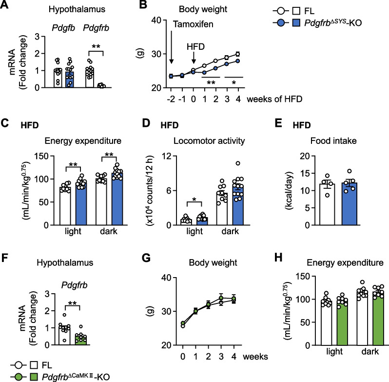
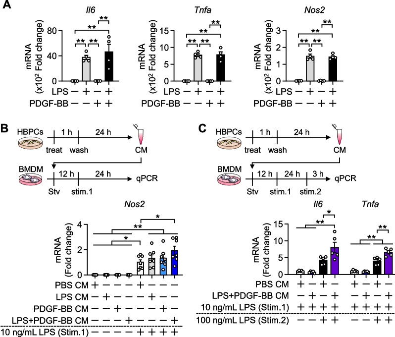
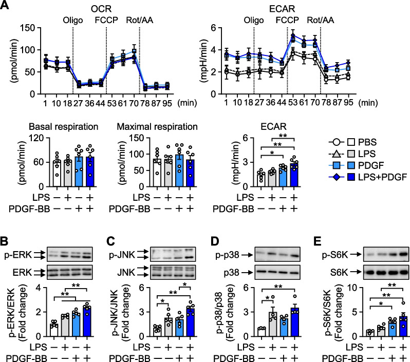
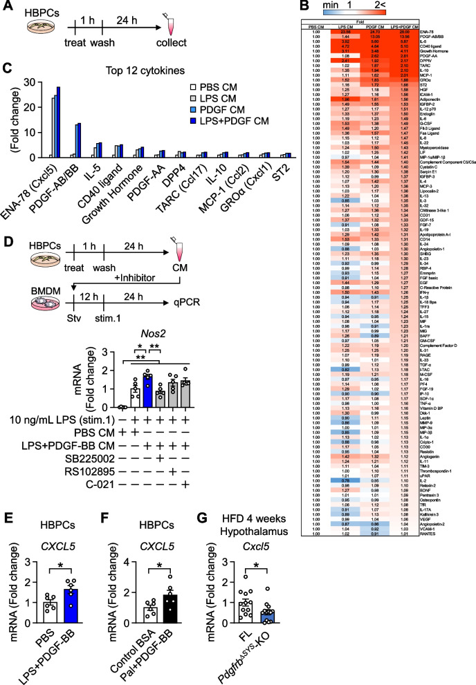
Methods: Tamoxifen-inducible systemic conditional PDGF receptor β knockout mice (Pdgfrb∆SYS-KO) and Calcium/calmodulin-dependent protein kinase type IIa (CaMKIIa)-positive neuron-specific PDGF receptor β knockout mice (Pdgfrb∆CaMKII-KO) were fed a high-fat diet, and metabolic phenotypes before and 3 to 4 weeks after dietary loading were examined. Intracellular energy metabolism and relevant signal transduction in lipopolysaccharide- and/or platelet-derived growth factor-BB (PDGF-BB)-stimulated human brain pericytes (HBPCs) were assessed by the Seahorse XFe24 Analyzer and Western blotting. The pericyte secretome in conditioned medium from HBPCs was studied using cytokine array kit, and its impact on polarization was examined in bone marrow-derived macrophages (BMDMs), which are microglia-like cells.

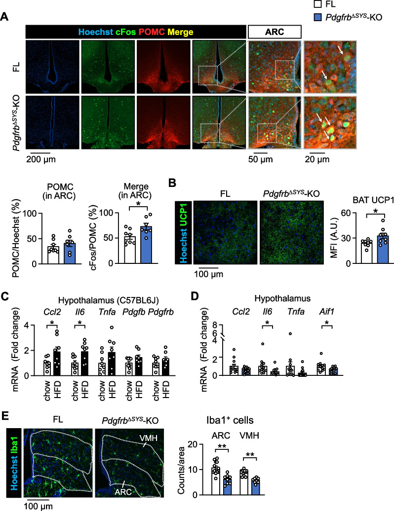
Results: Energy consumption increased and body weight gain decreased after high-fat diet loading in Pdgfrb∆SYS-KO mice. Cellular oncogene fos (cFos) expression increased in proopiomelanocortin (POMC) neurons, whereas microglial numbers and inflammatory gene expression decreased in the hypothalamus of Pdgfrb∆SYS-KO mice. No significant changes were observed in Pdgfrb∆CaMKII-KO mice. In HBPCs, a co-stimulation with lipopolysaccharide and PDGF-BB shifted intracellular metabolism towards glycolysis, activated mitogen-activated protein kinase (MAPK), and modulated the secretome to the inflammatory phenotype. Consequently, the secretome showed an increase in various proinflammatory chemokines and growth factors including Epithelial-derived neutrophil-activating peptide 78 (C-X-C motif chemokine ligand (CXCL)5), Thymus and activation-regulated chemokine (C-C motif chemokine (CCL)17), Monocyte chemoattractant protein 1 (CCL2), and Growth-regulated oncogene α (CXCL1). Furthermore, conditioned medium from HBPCs stimulated the inflammatory priming of BMDMs, and this change was abolished by the C-X-C motif chemokine receptor (CXCR) inhibitor. Consistently, mRNA expression of CXCL5 was elevated by lipopolysaccharide and PDGF-BB treatment in HBPCs, and the expression was significantly lower in the hypothalamus of Pdgfrb∆SYS-KO mice than in control Pdgfrbflox/flox mice (FL) following 4 weeks of HFD feeding.

Conclusions: PDGF receptor β signaling in hypothalamic pericytes promotes polarization of macrophages by changing their secretome and contributes to the progression of obesity.

Keywords: Inflammation; Microglia; Obesity; Pericytes; Platelet-derived growth factor.


Conflict of interest statement

The authors declare no competing interests.

Figures

Fig. 1
Fig. 1
PDGF signaling in pericytes, but not in neurons, mediates the dysfunction of energy metabolism. A Pdgfb and Pdgfrb mRNA levels in the hypothalamus of Pdgfrb∆SYS-KO fed HFD for 4 weeks (n = 12–13). B Body weights of Pdgfrb∆SYS-KO (n = 12–13). C–E Metabolic parameters of Pdgfrb∆SYS-KO fed HFD for 3–4 weeks. Energy expenditure (C), locomotor activity (D) (n = 9–12), and food intake (E) (n = 4–5). F Pdgfrb mRNA levels in the hypothalamus of Pdgfrb∆CaMKII-KO fed HFD for 4 weeks (n = 8–10). G Body weights of Pdgfrb∆CaMKII-KO (n = 8–10). H Energy expenditure in Pdgfrb∆CaMKII-KO fed HFD for 3 weeks (n = 8–10). Data are presented as the mean ± SEM. *p < 0.05 and **p < 0.01
Fig. 2
Fig. 2
PDGF signaling mediates chronic inflammation in the hypothalamus. A Representative confocal images of cFos (green) and POMC (red) immunoreactivities and counterstaining with Hoechst 33342 (blue) in the ARC of FL and Pdgfrb∆SYS-KO fed HFD for 4 weeks (n = 7–8). Scale bar = 200 μm. The right two panels are high magnification of the gray line square area. White arrows indicate Hoechst+cFos+POMC+ cells. Scale bar = 50 or 20 μm. The ratio of POMC-positive neurons in Hoechst-positive cells and cFos-POMC double-positive neurons in POMC-positive neurons are quantified. B Representative confocal images of UCP1 immunoreactivity (green) and Hoechst 33,342 (blue) staining in the BAT of FL and Pdgfrb∆SYS-KO fed HFD for 4 weeks. Scale bar = 100 μm. Mean fluorescent intensity (MFI) of UCP1 in the BAT (n = 8). C mRNA levels in the hypothalamus of C57BL6J mice fed a normal chow diet or HFD for 4 weeks (n = 8). D mRNA levels in the hypothalamus of FL and Pdgfrb∆SYS-KO fed HFD for 4 weeks (n = 12–13). E Representative confocal images of Iba1 immunoreactivity (green) in the ARC and VMH of FL and Pdgfrb∆SYS-KO fed HFD for 4 weeks (n = 8–12). Scale bar = 100 μm. Each region was indicated by a dotted line. The numbers of Iba1-positive microglia in the ARC and VMH were quantified. Data are presented as the mean ± SEM. *p < 0.05 and **p < 0.01
Fig. 3
Fig. 3
HBPC-derived conditioned medium induces the inflammatory polarization of BMDMs. A Il6, Tnfa, and Nos2 mRNA levels in BMDMs treated with 100 ng/mL LPS and/or 100 ng/mL recombinant human PDGF-BB for 3 h (n = 4). B Upper; timeline of the experiment. Lower; Nos2 mRNA level in BMDMs (n = 7). HBPCs were treated with LPS and/or PDGF-BB for 1 h, and cells were washed and replaced with fresh medium. The conditioned medium (CM) of HBPCs was harvested after 24 h. BMDMs were then treated with each CM in serum-starved medium for 12 h and primed with 10 ng/mL LPS (stim. 1) for 24 h. C Upper; timeline of the experiment. Lower; Il6 and Tnfa mRNA levels in BMDMs (n = 5). BMDMs were treated with each CM in serum-starved medium for 12 h, primed with 10 ng/mL LPS (stim. 1) for 24 h, and stimulated with 100 ng/mL LPS (stim. 2). Data are presented as the mean ± SEM. *p < 0.05 and **p < 0.01, significantly different between each group. Stv starvation, stim. stimulation
Fig. 4
Fig. 4
PDGF signaling shifts cellular metabolism towards glycolysis. A HBPCs were stimulated with LPS and/or PDGF-BB for 1 h and the oxygen consumption rate (OCR) and extracellular acidification rate (ECAR) were analyzed by a flux analyzer (n = 6). Oligo; oligomycin, FCCP; carbonyl cyanide-4 (trifluoromethoxy) phenylhydrazone, Rot/AA; a mix of rotenone/antimycin A. B–E HBPCs were treated with 100 ng/mL LPS and/or 100 ng/mL recombinant human PDGF-BB for 3 h. Representative Western blotting and quantified results are shown (n = 4–5). Data are presented as the mean ± SEM. *p < 0.05 and **p < 0.01, significantly different between each group
Fig. 5
Fig. 5
Secretome profile of HBPCs. A Timeline of CM collection. HBPCs were treated with LPS and/or PDGF-BB for 1 h, washed, cultured for 24 h, and CMs were collected. B Heat map of different protein levels in HBPC CM. The secreted levels of each factor are expressed as fold changes from the amount secreted in PBS CM. Colors from blue to red indicate a low to high protein level. C The top 12 proteins secreted in response to the stimulation are summarized in the graph. D Effects of inhibitors on the inflammatory priming of BMDMs by the pericyte secretome. BMDMs were pre-incubated with 1 μM SB225002 (CXCR1/2 inhibitor), 5 μM RS102895 (CCR2 inhibitor), or 100 nM C021 (CCR4 inhibitor) in serum-free RPMI 1640 media for 1 h, followed by a treatment with PBS CM or LPS + PDGF-BB CM for 12 h. Cells were then stimulated with 10 ng/mL LPS for 24 h, and Nos2 expression was measured as an index of inflammatory priming (n = 5). E mRNA level of CXCL5 in HBPCs treated with 100 ng/mL LPS and 100 ng/mL recombinant human PDGF-BB for 3 h (n = 6). F mRNA level of CXCL5 in HBPCs treated with 100 μM Palmitate (Pal) and 100 ng/mL recombinant human PDGF-BB for 6 h (n = 6). G Cxcl5 mRNA expression in the hypothalamus of FL and Pdgfrb∆SYS-KO fed HFD for 4 weeks (n = 12–13). Data are presented as the mean ± SEM. *p < 0.05 and **p < 0.01, significantly different between each group
Fig. 6
Fig. 6
Overview of the mechanism underlying chronic hypothalamic inflammation by pericyte-microglial interactions in early obesity. A presumed mechanism of obesity based on the present study. Upon HFD, pericytes receive inflammatory inputs in addition to PDGF signaling, which changes their secretome to an inflammatory phenotype. Secreted factors mobilize microglial polarization and enhance chronic inflammation in the hypothalamus. Inflammation modulates neuronal activity and decreases energy expenditure to further promote obesity

Similar articles

Cited by

References

    1. Alluri H, Wilson RL, Anasooya Shaji C, Wiggins-Dohlvik K, Patel S, et al. Melatonin preserves blood-brain barrier integrity and permeability via matrix metalloproteinase-9 Inhibition. PLoS ONE. 2016;11(5):e0154427. doi: 10.1371/journal.pone.0154427. - DOI - PMC - PubMed
    1. Amar J, Burcelin R, Ruidavets JB, Cani PD, Fauvel J, et al. Energy intake is associated with endotoxemia in apparently healthy men. Am J Clin Nutr. 2008;87(5):1219–1223. doi: 10.1093/ajcn/87.5.1219. - DOI - PubMed
    1. Biglari N, Gaziano I, Schumacher J, Radermacher J, Paeger L, et al. Functionally distinct POMC-expressing neuron subpopulations in hypothalamus revealed by intersectional targeting. Nat Neurosci. 2021;24(7):913–929. doi: 10.1038/s41593-021-00854-0. - DOI - PMC - PubMed
    1. Boro M, Balaji KN. CXCL1 and CXCL2 regulate NLRP3 inflammasome activation via G-protein-coupled receptor CXCR2. J Immunol. 2017;199(5):1660–1671. doi: 10.4049/jimmunol.1700129. - DOI - PubMed
    1. Brown LS, Foster CG, Courtney JM, King NE, Howells DW, et al. Pericytes and neurovascular function in the healthy and diseased brain. Front Cell Neurosci. 2019;13:282. doi: 10.3389/fncel.2019.00282. - DOI - PMC - PubMed

MeSH terms

Substances