. 2024 Feb 5;26(1):23.
doi: 10.1186/s13058-024-01778-w.

The novel phosphatase NUDT5 is a critical regulator of triple-negative breast cancer growth

Affiliations

The novel phosphatase NUDT5 is a critical regulator of triple-negative breast cancer growth

Jing Qian et al. Breast Cancer Res. .

Erratum in

Abstract

Background: The most aggressive form of breast cancer is triple-negative breast cancer (TNBC), which lacks expression of the estrogen receptor (ER) and progesterone receptor (PR), and does not have overexpression of the human epidermal growth factor receptor 2 (HER2). Treatment options for women with TNBC tumors are limited, unlike those with ER-positive tumors that can be treated with hormone therapy, or those with HER2-positive tumors that can be treated with anti-HER2 therapy. Therefore, we have sought to identify novel targeted therapies for TNBC. In this study, we investigated the potential of a novel phosphatase, NUDT5, as a potential therapeutic target for TNBC.

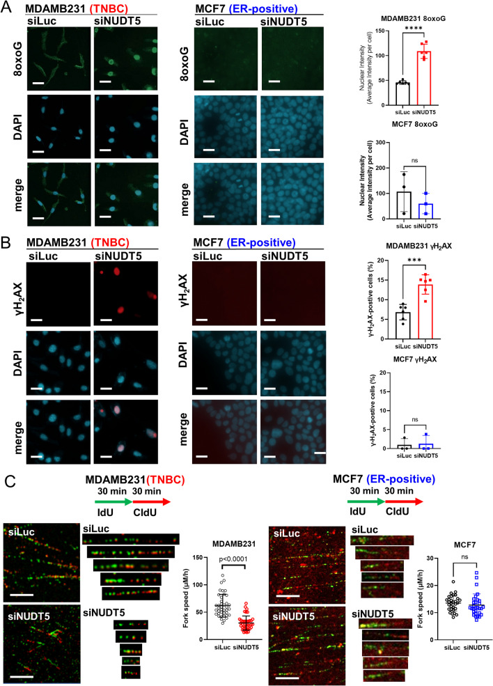
Methods: The mRNA expression levels of NUDT5 in breast cancers were investigated using TCGA and METABRIC (Curtis) datasets. NUDT5 ablation was achieved through siRNA targeting and NUDT5 inhibition with the small molecule inhibitor TH5427. Xenograft TNBC animal models were employed to assess the effect of NUDT5 inhibition on in vivo tumor growth. Proliferation, death, and DNA replication assays were conducted to investigate the cellular biological effects of NUDT5 loss or inhibition. The accumulation of 8-oxo-guanine (8-oxoG) and the induction of γH2AX after NUDT5 loss was determined by immunofluorescence staining. The impact of NUDT5 loss on replication fork was assessed by measuring DNA fiber length.

Results: In this study, we demonstrated the significant role of an overexpressed phosphatase, NUDT5, in regulating oxidative DNA damage in TNBCs. Our findings indicate that loss of NUDT5 results in suppressed growth of TNBC both in vitro and in vivo. This growth inhibition is not attributed to cell death, but rather to the suppression of proliferation. The loss or inhibition of NUDT5 led to an increase in the oxidative DNA lesion 8-oxoG, and triggered the DNA damage response in the nucleus. The interference with DNA replication ultimately inhibited proliferation.

Conclusions: NUDT5 plays a crucial role in preventing oxidative DNA damage in TNBC cells. The loss or inhibition of NUDT5 significantly suppresses the growth of TNBCs. These biological and mechanistic studies provide the groundwork for future research and the potential development of NUDT5 inhibitors as a promising therapeutic approach for TNBC patients.

Keywords: Oxidative stress; Phosphatase; Triple-negative breast cancer.


Conflict of interest statement

P.H.B served as a Scientific Advisory Board Member for the Susan G. Komen for the Cure Foundation (until 2017), and P.H.B is a holder of GeneTex stock (less than 1% of the total company stock); neither of these relate to this publication. All remaining authors declare no actual, potential, or perceived conflict of interest that would prejudice the impartiality of this article.

Figures

Fig. 1
Fig. 1
NUDT5 expression in breast cancer. A NUDT5 mRNA expression levels from publicly available databases (from The Cancer Genome Atlas and Molecular Taxonomy of Breast Cancer International Consortium [METABRIC]). The mRNA expression level of NUDT5 is compared between TNBC and ER-positive breast cancers, as well as TNBC and normal breast. B NUDT5 mRNA expression levels across the different PAM50 subgroups using the METABRIC data. The mRNA expression level of NUDT5 is compared between basal subtype and the normal-like, Luminal A, Luminal B, HER2-enriched, and Claudin-low subtypes. C NUDT5 expression across the different triple-negative breast cancer (TNBC) subtypes (from Burstein et al. [32]) using data from METABRIC. D NUDT5 mRNA expression in non-TNBC and TNBC cell lines using data from the Cancer Cell Line Encyclopedia is shown [25]. E NUDT5 protein expression is shown by Western blot analysis of multiple cell lines, including normal-like, estrogen receptor-positive, and TNBC cell lines. Quantification of NUDT5 protein levels in non-TNBC and TNBC cells is also shown. The differences of NUDT5 expression levels between two subtypes were determined by Student’s t test and the differences among multiple subtypes were determined by one-way ANOVA. (ns not significant; *p < 0.05; **p < 0.01; ***p < 0.001; ****p < 0.0001)
Fig. 2
Fig. 2
NUDT5 loss inhibits triple-negative breast cancer growth. A Cell growth of estrogen receptor-positive breast cancer cells (MCF7, ZR-75-1 and MDA-MB-361), and triple-negative breast cancer cells (MDA-MB-231, MDA-MB-436, and MDA-MB-468) following siLuc or siNUDT5 treatment. Knockdown efficiency of protein samples harvested at Day 7 is shown by Western blot analysis (protein expression at D1 and D7 is also shown in Additional file 2: Figure S2A). RNA expression after siRNA knockdown at D1 and D7 is also shown in Additional file 2: Figure S2A. The significant differences between day 7 cell counts were determined using Student’s t test (ns not significant; *p < 0.05; **p < 0.01; ***p < 0.001; ****p < 0.0001). Shown in this figure is one representative growth experiment for each cell line. This experiment was repeated in each of these cell lines with similar results showing effective siRNA knockdown (at the RNA and protein levels), as well as effective growth suppression in the TNBC cell lines
Fig. 3
Fig. 3
NUDT5 small molecule inhibitor TH5427 suppresses triple-negative breast cancer growth in vitro and in vivo. A ER-positive cell lines MCF7 and ZR-75-1 and TNBC cell lines MDA-MB-231 and MDA-MB-436 were treated with DMSO control or 10 μM TH5427 at day 1. Cell counts were recorded on days 1, 3, 5, and 7. B Schematic illustration of MDA-MB-231 xenograft tumors. 20 Nude mice were injected with MDA-MB-231 cells, and randomized into two groups of 10 nude mice each, which were treated with either vehicle or 50 mg/kg TH5427 via intraperitoneal injection 5 times per week. Mice were sacrificed when the largest tumor size reached 1000 mm3. 4 mice were found dead after 7 days of TH5427 treatment. Tumor volumes were calculated using the formula (width × width × length)/2. Tumor growth was analyzed using linear regression of log10 (tumor volume), and the difference between tumor growth slopes was compared by Student’s t test (*p < 0.05; ***p < 0.001). C H&E and IHC staining of tumor samples from 20 female nude mice injected with MDA-MB-231 cells into the mammary fat pad. H&E and IHC images of one representative tumor are shown. Additional images from 4 other tumors (2 tumors from vehicle-treated mice and 2 tumors from TH5427-treated mice) are presented in Additional file 3: Figure S3C. Ki67 positivity was compared between treatment and control groups. Also see Additional file 3: Figure S3D of the NUDT5 expression via IHC analysis of the 6 independent tumors outlined in 3C above (3 tumors from vehicle-treated mice and 3 tumors from TH55427-treated mice). NUDT5 expression is also shown via IHC analysis of MDA-MD-361 cells (negative control) and MDA-MB-231 cells (positive control). The statistical difference in these analyses was determined by Student’s t test
Fig. 4
Fig. 4
NUDT5 depletion suppresses TNBC cell proliferation. A BrdU incorporation in the ER-positive cell line MCF-7 and TNBC cell line MDA-MB-231 treated with siLuc versus siNUDT5, or DMSO versus TH5427. Cells were treated 48 h prior to the assay. B DRAQ7 cell death assay in the ER-positive cell line MCF-7 and TNBC cell line MDA-MB-231 treated with siLuc versus siNUDT5, or DMSO versus TH5427. Cells were treated 48 h prior to the assay. 10 µM staurosporine was used as a positive control. C Annexin V/ propidium iodide (PI) staining in the ER-positive cell line MCF-7 and TNBC cell line MDA-MB-231 treated with siLuc versus siNUDT5, or DMSO versus TH5427. Cells were treated 48 h prior to the assay. 1 µM bortezomib or 10 µM staurosporine were used as a positive control. The apoptotic cell population is composed of both early (Annexin V-positive/PI-negative) and late apoptotic (Annexin V-positive/PI-positive) cells. Statistical comparisons were analyzed by the Student’s t test, and p values are shown
Fig. 5
Fig. 5
Loss of NUDT5 induces oxidative 8-oxoG and DNA damage response. A 8-oxoG lesions were stained in TNBC (MDA-MB-231) and ER-positive (MCF-7) cells treated with siLuc or siNUDT5, and nuclei were counterstained with DAPI after 4 days. The data is shown as nuclear intensity for siLuc- or siNUDT5-treated cells. B γH2AX was stained in MDA-MB-231 and MCF-7 cells treated with siLuc or siNUDT5, and nuclei were counterstained with DAPI after 7 days. The data is shown as γH2AX positivity, and was compared between the different treatments. Statistical significance was analyzed by the Student’s t test. C Representative DNA fibers from MDA-MB-231 and MCF-7 cells treated with siLuc or siNUDT5. Cells were first labelled with 30 min treatment with 50 µM IdU, followed by 30 min treatment with 100 µM CIdU. DNA fiber length was imaged by an Andor Revolution XDi WD Spinning disk confocal microscope and analyzed with Imaris software. The fork speed = (the length of IdU labeled DNA fiber + the length of CdU labeled DNA fiber)/total labeling time (1 h). The difference between siLuc- and siNDUT5-treated DNA fibers is shown graphically and compared using a Student’s t test. Additional TNBC (MDA-MB-436, MDA-MB-468) and ER-positive (ZR-75-1 and MDA-MB-361) cell lines are shown in Additional file 7, 8: Figures S7 and S8. Proof of effective knockdown is shown via Western blot and qPCR in Additional file 2: Figure S2A
Fig. 6
Fig. 6
Proposed mechanism of NUDT5 biological function in breast cancers. The level of ROS is low in ER-positive tumors, resulting in low accumulation of 8-oxoG and γH2AX lesions in the nucleus. In such cases, the inhibition or loss of NUDT5 does not affect the growth of these ER-positive tumors. In TNBC tumors, both ROS and NUDT5 levels are elevated. When NUDT5 is abundant, it mitigates oxidative DNA damage by hydrolyzing oxidized deoxyribonucleoside diphosphates. Consequently, there is no incorporation of 8-oxoG into the DNA, and γH2AX lesions do not accumulate. However, in the absence of NUDT5, uncontrolled oxidative stress on the nucleotide pool occurs. This leads to the incorporation of 8-oxoG lesions into the DNA and the accumulation of γH2AX lesions in the nucleus, ultimately causing DNA replication fork slowing and reduced proliferation

References

    1. Sung H, Ferlay J, Siegel RL, Laversanne M, Soerjomataram I, Jemal A, et al. Global cancer statistics 2020: GLOBOCAN estimates of incidence and mortality worldwide for 36 cancers in 185 countries. CA Cancer J Clin. 2021;71(3):209–249. doi: 10.3322/caac.21660. - DOI - PubMed
    1. Siegel RL, Miller KD, Fuchs HE, Jemal A. Cancer statistics, 2022. CA Cancer J Clin. 2022;72(1):7–33. doi: 10.3322/caac.21708. - DOI - PubMed
    1. Waks AG, Winer EP. Breast cancer treatment. JAMA. 2019;321(3):288–300. doi: 10.1001/jama.2018.19323. - DOI - PubMed
    1. Bianchini G, De Angelis C, Licata L, Gianni L. Treatment landscape of triple-negative breast cancer—expanded options, evolving needs. Nat Rev Clin Oncol. 2022;19(2):91–113. doi: 10.1038/s41571-021-00565-2. - DOI - PubMed
    1. Speers C, Tsimelzon A, Sexton K, Herrick AM, Gutierrez C, Culhane A, et al. Identification of novel kinase targets for the treatment of estrogen receptor-negative breast cancer. Clin Cancer Res. 2009;15(20):6327–6340. doi: 10.1158/1078-0432.CCR-09-1107. - DOI - PMC - PubMed

Publication types