

Investigation of the Effect of Seminal Plasma Exosomes from the Normal and Oligoasthenoteratospermic Males in the Implantation Process
- PMID: 38317811
- PMCID: PMC10838591
- DOI: 10.61186/rbmb.12.2.294
Investigation of the Effect of Seminal Plasma Exosomes from the Normal and Oligoasthenoteratospermic Males in the Implantation Process
Abstract
Background: Seminal plasma exosomes are now recognized to play a complex role in the regulation of the female reproductive system infertility. The objective of this study was to assess the effect of exosomes derived from the sperm of men with oligoasthenoteratozoospermia on endometrial implantation-related genes.
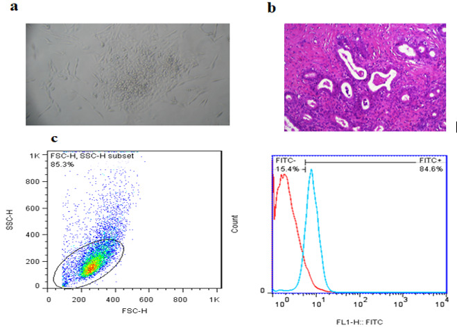
Methods: To isolate the exosomes, we employed an ultracentrifugation method on samples derived from 10 fertile men with normal sperm parameters and 10 men with oligoasthenoteratozoospermia. The size distribution and ultrastructure of the exosomes were then characterized using transmission electron microscopy and dynamic light scattering. We detected an exosome marker using western blot analysis and confirmed the cytoplasmic localization of the exosomes by incubating them with DiI dye and visualizing them using fluorescence microscopy. After 6 hours of in vitro treatment of endometrial epithelial cells with 100 µg/ml seminal exosome, the endometrial receptivity genes were examined using qRT-PCR. To perform data analysis and quantification, we utilized Image J and Prism software. P< 0.05 were considered statistically significant.
Results: After 6 hours of treatment, the mRNA levels of MUC1, LIF, G-CSF, CX3CL1, and VEGF were significantly downregulated in the endometrial epithelial cells treated with oligoasthenoteratozoospermia exosomes compared to the normal group. Although changes were observed in the mean mRNA levels of IL8 and TGF-β genes in the oligoasthenoteratozoospermia group compared to the normal group, these differences did not reach statistical significance (p > 0.05).
Conclusions: Oligoasthenoteratozoospermia exosomes have a distinct effect on endometrial receptivity compared to normal exosomes, leading to reduced expression of implantation-related genes.
Keywords: Embryo implantation; Endometrium; Exosome; Infertility; Semen.
Conflict of interest statement
The authors declare no conflicts of interest.
Figures


Similar articles
-
[The miR-184 level in the seminal plasma exosome of male infertility patients and its clinical significance].Zhonghua Nan Ke Xue. 2020 Aug;26(8):686-694. Zhonghua Nan Ke Xue. 2020. PMID: 33377728 Chinese.
-
A human chorionic gonadotropin (hCG) delivery platform using engineered uterine exosomes to improve endometrial receptivity.Life Sci. 2021 Jun 15;275:119351. doi: 10.1016/j.lfs.2021.119351. Epub 2021 Mar 15. Life Sci. 2021. PMID: 33737084
-
Seminal exosomes induce interleukin-6 and interleukin-8 secretion by human endometrial stromal cells.Eur J Obstet Gynecol Reprod Biol. 2019 Apr;235:71-76. doi: 10.1016/j.ejogrb.2019.02.010. Epub 2019 Feb 19. Eur J Obstet Gynecol Reprod Biol. 2019. PMID: 30807994
-
Review: Embryo- and endometrium-derived exosomes and their potential role in assisted reproductive treatments-liquid biopsies for endometrial receptivity.Placenta. 2017 Jun;54:89-94. doi: 10.1016/j.placenta.2016.12.011. Epub 2016 Dec 9. Placenta. 2017. PMID: 28043656 Review.
-
Potential roles of metalloproteinases of endometrium-derived exosomes in embryo-maternal crosstalk during implantation.J Cell Physiol. 2018 Jun;233(6):4530-4545. doi: 10.1002/jcp.26259. Epub 2017 Dec 26. J Cell Physiol. 2018. PMID: 29115666 Review.
Cited by
-
Seminal Plasma Extracellular Vesicles: Key Mediators of Intercellular Communication in Mammalian Reproductive Systems.Vet Sci. 2025 Jun 13;12(6):585. doi: 10.3390/vetsci12060585. Vet Sci. 2025. PMID: 40559822 Free PMC article. Review.
References
-
- Barratt CLR, Wang C, Baldi E, Toskin I, Kiarie J, Lamb DJ. What advances may the future bring to the diagnosis, treatment, and care of male sexual and reproductive health? Fertil Steril. 2022;117(2):258–267. other Editorial Board Members of the WHO Laboratory Manual for the Examination and Processing of Human Semen. - PMC - PubMed
-
- Palermo G, Joris H, Devroey P, Van Steirteghem AC. Pregnancies after intracytoplasmic injection of single spermatozoon into an oocyte. Lancet. 1992;340(8810):17–8. - PubMed
-
- Mohamed Khosroshahi L, Parhizkar F, Kachalaki S, Aghebati-Maleki A, Aghebati-Maleki L. Immune checkpoints and reproductive immunology: Pioneers in the future therapy of infertility related Disorders? Int Immunopharmacol. 2021;99:107935. - PubMed
-
- Schjenken JE, Glynn DJ, Sharkey DJ, Robertson SA. TLR4 Signaling Is a Major Mediator of the Female Tract Response to Seminal Fluid in Mice. Biol Reprod. 2015;93(3):68. - PubMed
LinkOut - more resources
Full Text Sources
Research Materials
Miscellaneous