. 2024 Jan 22:12:1338419.
doi: 10.3389/fcell.2024.1338419. eCollection 2024.

C5L2 CRISPR KO enhances dental pulp stem cell-mediated dentinogenesis via TrkB under TNFα-induced inflammation

Affiliations

C5L2 CRISPR KO enhances dental pulp stem cell-mediated dentinogenesis via TrkB under TNFα-induced inflammation

Muhammad Irfan et al. Front Cell Dev Biol. .

Abstract

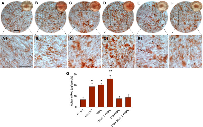
Background and Objectives: Dental caries is one of the most common human pathological conditions resulting from the invasion of bacteria into the dentin. Current treatment options are limited. In many cases, endodontic therapy leads to permanent pulp tissue loss. Dentin-pulp complex regeneration involves dental pulp stem cells (DPSCs) that differentiate into odontoblast-like cells under an inflammatory context. However, limited information is available on how DPSC differentiation processes are affected under inflammatory environments. We identified the crucial role of complement C5a and its receptor C5aR in the inflammation-induced odontoblastic DPSC differentiation. Methodology: Here, we further investigated the role of a second and controversial C5a receptor, C5L2, in this process and explored the underlying mechanism. Human DPSCs were examined during 7-, 10-, and 14-day odontogenic differentiation treated with TNFα, C5L2 CRISPR, and tyrosine receptor kinase B (TrkB) antagonist [cyclotraxin-B (CTX-B)]. Results: Our data demonstrate that C5L2 CRISPR knockout (KO) enhances mineralization in TNFα-stimulated differentiating DPSCs. We further confirmed that C5L2 CRISPR KO significantly enhances dentin sialophosphoprotein (DSPP) and dentin matrix protein-1 (DMP-1) expression after 14-day odontoblastic DPSC differentiation, and treatment with CTX-B abolished the TNFα/C5L2 CRISPR KO-induced DSPP and DMP-1 increase, suggesting TrkB's critical role in this process. Conclusion and Key applications: Our data suggest a regulatory role of C5L2 and TrkB in the TNFα-induced odontogenic DPSC differentiation. This study may provide a useful tool to understand the mechanisms of the role of inflammation in dentinogenesis that is required for successful DPSC engineering strategies.

Keywords: C5L2; DPSC; TrkB; complement C5a; dentinogenesis; inflammation.


Conflict of interest statement

The authors declare that the research was conducted in the absence of any commercial or financial relationships that could be construed as a potential conflict of interest.

Figures

FIGURE 1
FIGURE 1
Differentiating DPSCs express C5L2, and TNFα stimulation enhances its expression. (A) Experimental scheme showing various stages of differentiation and treatments. (B) Commercial DPSCs were further confirmed with a mesenchymal stem cell marker STRO-1. Nuclei were counter-stained with DAPI. (C) DPSCs constitutively express C5L2. (D) TNFα stimulation enhances the expression of C5L2 [scale bar (B–D): 50 μm]. (E–J) Cultured DPSCs showing various stages of differentiation in dentinogenic media under a light microscope (scale bar: 200 μm) or phase contrast microscopy [scale bar (G, J): 50 μm]. (K) To evaluate whether various treatments affect the proliferation and differentiation rate, cell density was measured by cell counting, and no significant difference was found. The graph shows the mean ± SD of at least three independent experiments (n = 3) in duplicate. (L) To validate C5L2 CRISPR/Cas9 KO, a Western blotting assay was performed. C5L2 KO plasmid treated well clearly indicated C5L2 KO compared with control cells. (M) The expression of odontoblastic markers ALP, BMP-2, COL1A1, and DMP-1 mRNA during the odontogenic differentiation was quantified by real-time PCR. The elevated level of these markers represents the odontoblast-like differentiation of DPSCs. *p < 0.05 and **p < 0.01 vs. day 0-fold change. The bar graph shows the mean ± SD of at least three independent experiments (n = 3) in duplicate.
FIGURE 2
FIGURE 2
C5L2 KO DPSCs secrete more mineralized substances during odontoblastic differentiation. Control cells (A–D) show less mineralized substances than the C5L2 KO group (E–H) after 10 days of differentiation in dentinogenic media, while TNFα stimulation further enhanced the mineralization matrix (I–L). (M–P) Images show calcium crystals in various treatment groups of differentiating DPSCs. TNFα-treated odontoblastic differentiating C5L2 KO DPSCs secrete more calcium crystals than C5L2 KO or TNFα alone compared with the control. The results shown are indicative of at least three independent experiments (n = 3) in duplicate.
FIGURE 3
FIGURE 3
Mineralization activity of differentiated C5L2 KO DPSCs in dentinogenic media. (A–F1) Differentiated cells were stained with ARS, and images were taken using a Leica DMi1 phase microscope. Images show higher mineralization matrix in the C5L2 KO (B, B1) or TNFα alone (C, C1) or their combined treated group (D, D1) compared with the control (A, A1), while the effects were reversed in the CTX-treated groups (E–F1). Scale bars: 100 μm. (G) Bar graph shows ARS quantification among various treatment groups indicating significantly higher peaks in the C5L2 KO, TNFα, or their combined treatment, while CTX treatment reversed their effects. The graph shows the mean ± SD of at least three independent experiments (n = 3) in duplicate. *p < 0.05 and **p < 0.01 vs. control.
FIGURE 4
FIGURE 4
Effects of C5L2 KO on mineralization markers DSPP and DMP-1. (A–X) Double fluorescent immunostaining was performed (DSPP: green; DMP-1: red) after 10 days of DPSC odontoblastic differentiation in dentinogenic media with or without TNFα or CTX in C5L2 KO cells. Cells were counter-stained with DAPI (blue). (A–D) Control cells showed less DSPP and DMP-1 expression than the C5L2 KO (E–H) or TNFα alone (I–L)-treated groups, while their combined effect is significantly higher (M–P). CTX treatment significantly abolished the effect of C5L2 KO or TNFα (Q–X). (Y) The bar graph shows a significant increment in DSPP and DMP-1 expression, and even more enhanced expression in the combined treated group was reversed by CTX treatment. The graph shows the mean ± SD of at least three independent experiments (n = 3) in duplicate. *p < 0.05 and ***p < 0.001 vs. control. ### p < 0.001 vs. C5L2 KO and TNFα-combined group (Z). Line graph showing the co-localization of DSPP and DMP-1 being the highest peaks observed in C5L2 KO and TNFα-treated groups.
FIGURE 5
FIGURE 5
In-cell Western assay showing the expression of DSPP and DMP-1 in various treatment groups with or without TNFα or CTX in the C5L2 KO or control odontoblastic differentiated DPSCs. (A, B) Cells were assayed by the in-cell Western technique and photographed using Odyssey CLx; C5L2 KO and TNFα showed higher intensities under red and green channels against beta-actin. (A1–B1) 2.5D model shows the highest peaks among the C5L2 KO and TNFα alone or combined treated group. (C) The bar graph shows the increased expression of DSPP and DMP-1 in C5L2 KO and TNFα-treated groups. The graph shows the mean ± SD of at least three independent experiments (n = 3) in duplicate. *p < 0.05 and ***p < 0.001 vs. control. ## p < 0.01 vs. C5L2 KO and TNFα-combined groups.
FIGURE 6
FIGURE 6
Effects of C5L2 KO or TNFα on the secretion of DSPP and DMP-1 in supernatants of differentiated DPSCs. (A, B) Cells were cultured and incubated with various treatments such as C5L2 KO plasmid, TNFα alone, or combined with CTX. The supernatants (conditioned media) were collected at days 7, 10, and 14 during differentiation, and an ELISA was performed to quantify DSPP and DMP-1 secretion in C5L2-mediated DPSCs according to the manufacturer’s protocol. (A) In the supernatant, DSPP and DMP-1 production was significantly increased in the C5L2 KO cells with or without TNFα at all stages. However, CTX treatment reversed the DSPP and DMP-1 production. The graph shows the mean ± SD of at least three independent experiments (n = 3) in duplicate. *p < 0.05, **p < 0.01, and ***p < 0.001 vs. control; ###p < 0.001 vs. the respective line-indicated group.
FIGURE 7
FIGURE 7
Summarized effects of C5L2 CRISPR/Cas9 KO on DPSCs.

References

    1. Abdel Meguid E., Ke Y., Ji J., El-Hashash A. H. K. (2018). Stem cells applications in bone and tooth repair and regeneration: new insights, tools, and hopes. J. Cell. physiology 233 (3), 1825–1835. 10.1002/jcp.25940 - DOI - PubMed
    1. About I. (2011). Dentin regeneration in vitro: the pivotal role of supportive cells. Adv. Dent. Res. 23 (3), 320–324. 10.1177/0022034511405324 - DOI - PubMed
    1. Bathina S., Das U. N. (2015). Brain-derived neurotrophic factor and its clinical implications. Archives Med. Sci. 11 (6), 1164–1178. 10.5114/aoms.2015.56342 - DOI - PMC - PubMed
    1. Bernar A., Gebetsberger J. V., Bauer M., Streif W., Schirmer M. (2022). Optimization of the Alizarin red S assay by enhancing mineralization of osteoblasts. Int. J. Mol. Sci. 24 (1), 723. 10.3390/ijms24010723 - DOI - PMC - PubMed
    1. Caplan A. I. (2007). Adult mesenchymal stem cells for tissue engineering versus regenerative medicine. J. Cell. physiology 213 (2), 341–347. 10.1002/jcp.21200 - DOI - PubMed