Paneth cell ontogeny in term and preterm ovine models
- PMID: 38318150
- PMCID: PMC10839032
- DOI: 10.3389/fvets.2024.1275293
Paneth cell ontogeny in term and preterm ovine models
Abstract
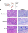
Introduction: Paneth cells are critically important to intestinal health, including protecting intestinal stem cells, shaping the intestinal microbiome, and regulating host immunity. Understanding Paneth cell biology in the immature intestine is often modeled in rodents with little information in larger mammals such as sheep. Previous studies have only established the distribution pattern of Paneth cells in healthy adult sheep. Our study aimed to examine the ontogeny, quantification, and localization of Paneth cells in fetal and newborn lambs at different gestational ages and with perinatal transient asphyxia. We hypothesized that ovine Paneth cell distribution at birth resembles the pattern seen in humans (highest concentrations in the ileum) and that ovine Paneth cell density is gestation-dependent.
Methods: Intestinal samples were obtained from 126-127 (preterm, with and without perinatal transient asphyxia) and 140-141 (term) days gestation sheep. Samples were quantified per crypt in at least 100 crypts per animal and confirmed as Paneth cells through in immunohistochemistry.
Results: Paneth cells had significantly higher density in the ileum compared to the jejunum and were absent in the colon.
Discussion: Exposure to perinatal transient asphyxia acutely decreased Paneth cell numbers. These novel data support the possibility of utilizing ovine models for understanding Paneth cell biology in the fetus and neonate.
Keywords: Paneth cell; immature intestine; ovine; perinatal bradycardic stress; preterm.
Copyright © 2024 Bautista, Cera, Schoenauer, Persiani, Lakshminrusimha, Chandrasekharan, Gugino, Underwood and McElroy.
Conflict of interest statement
SM is an advisor to LactaLogics, Evive, Noveome, and the NEC Society. The remaining authors declare that the research was conducted in the absence of any commercial or financial relationships that could be construed as a potential conflict of interest. The reviewer BS declared a past co-authorship with the author(s) SM to the handling editor.
Figures


Similar articles
-
The Paneth Cell: The Curator and Defender of the Immature Small Intestine.Front Immunol. 2020 Apr 3;11:587. doi: 10.3389/fimmu.2020.00587. eCollection 2020. Front Immunol. 2020. PMID: 32308658 Free PMC article. Review.
-
The stem-cell zone of the small intestinal epithelium. II. Evidence from paneth cells in the newborn mouse.Am J Anat. 1981 Jan;160(1):65-75. doi: 10.1002/aja.1001600106. Am J Anat. 1981. PMID: 7211717
-
Expression and Localization of Paneth Cells and Their α-Defensins in the Small Intestine of Adult Mouse.Front Immunol. 2020 Oct 13;11:570296. doi: 10.3389/fimmu.2020.570296. eCollection 2020. Front Immunol. 2020. PMID: 33154750 Free PMC article.
-
Characterization of paneth cells in alpacas (Vicugna pacos, Mammalia, Camelidae).Tissue Cell. 2016 Aug;48(4):383-8. doi: 10.1016/j.tice.2016.04.003. Epub 2016 May 7. Tissue Cell. 2016. PMID: 27233914 Free PMC article.
-
Paneth cells in intestinal physiology and pathophysiology.World J Gastrointest Pathophysiol. 2017 Nov 15;8(4):150-160. doi: 10.4291/wjgp.v8.i4.150. World J Gastrointest Pathophysiol. 2017. PMID: 29184701 Free PMC article. Review.

