Analysis of Variation in the Origin of the Obturator Artery in Midwestern American Donor Bodies
- PMID: 38318277
- PMCID: PMC10843247
- DOI: 10.7759/cureus.53650
Analysis of Variation in the Origin of the Obturator Artery in Midwestern American Donor Bodies
Abstract
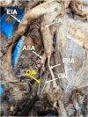
The obturator artery (OA) is typically a branch of the anterior division of the internal iliac artery. However, an aberrant obturator artery origin may lead to clinical complications. Because of its location in the pelvic cavity, the OA is at high risk of injury or laceration during a variety of pelvic surgeries. Regarding this, variations in the origins of the OA may result in bleeding that can often be overlooked, rendering treatment ineffective. Our study aimed to assess the origins and course of the OA in Midwestern American donor bodies. Sixty-two donor bodies were obtained from the Gift of Body Donation Program at A.T. Still University's Kirksville College of Osteopathic Medicine. The origin of each OA was documented and photographed. The OA was identified by observing the vessel's passage through the obturator foramen. Of 132 OAs studied, 72 (54.5%) had an aberrant OA. Further, 22 (16.7%) had an aberrant OA origin from the inferior epigastric artery, 20 (15.2%) had an aberrant OA origin from the posterior division of the internal iliac artery, 22 (16.7%) had an aberrant OA origin from dual origins of the anterior division of the internal iliac artery and the inferior epigastric artery, and eight (6.1%) had other aberrant OA origins. Overall, our results indicated anatomical variations are common in the origins and course of the OA. These data highlight the importance of considering variations in the OA and the prevalence of those variations during vascular and orthopedic procedures.
Keywords: anatomical variation; obturator artery; pelvic surgery; pelvic vasculature; pelvis.
Copyright © 2024, Diemer et al.
Conflict of interest statement
The authors have declared that no competing interests exist.
Figures


References
-
- Anatomical variations of corona mortis in the anterior intrapelvic approach: a cadaveric study. Sambhav K, Nayyar AK, Elhence A, Gupta R, Ghatak S. https://pubmed.ncbi.nlm.nih.gov/35780370/ Mymensingh Med J. 2022;31:826–834. - PubMed
-
- Variability of the obturator vessels. Gilroy AM, Hermey DC, DiBenedetto LM, Marks SC Jr, Page DW, Lei QF. https://pubmed.ncbi.nlm.nih.gov/9283731/ Clin Anat. 1997;10:328–332. - PubMed
-
- Aberrant obturator artery is a common arterial variant that may be a source of unidentified hemorrhage in pelvic fracture patients. Requarth JA, Miller PR. J Trauma. 2011;70:366–372. - PubMed
LinkOut - more resources
Full Text Sources
