

Central Autonomic Network and Heart Rate Variability in Premature Neonates
- PMID: 38320522
- PMCID: PMC11300706
- DOI: 10.1159/000536513
Central Autonomic Network and Heart Rate Variability in Premature Neonates
Abstract
Introduction: The Central Autonomic Network (CAN) is a hierarchy of brain structures that collectively influence cardiac autonomic input, mediating the majority of brain-heart interactions, but has never been studied in premature neonates. In this study, we use heart rate variability (HRV), which has been described as the "primary output" of the CAN, and resting-state functional MRI (rsfMRI) to characterize brain-heart relationships in premature neonates.
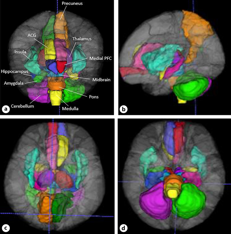
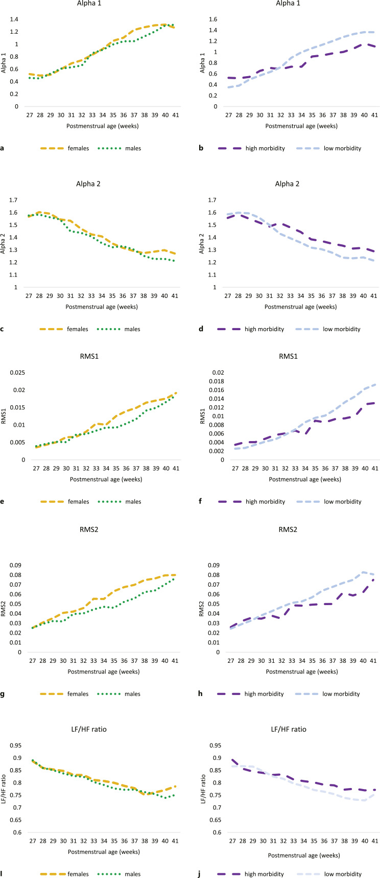
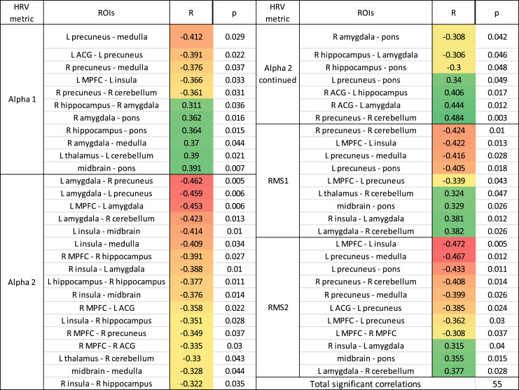
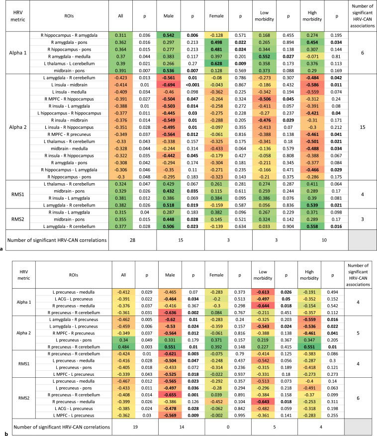
Methods: We studied premature neonates who underwent rsfMRI at term (37-week postmenstrual age or above) and had HRV data recorded during the same week of their MRI. HRV was derived from continuous electrocardiogram data during the week of the rsfMRI scan. For rsfMRI, a seed-based approach was used to define regions of interest (ROIs) pertinent to the CAN, and blood oxygen level-dependent signal was correlated between each ROI as a measure of functional connectivity. HRV was correlated with CAN connectivity (CANconn) for each region, and subgroup analysis was performed based on sex and clinical comorbidities.
Results: Forty-seven premature neonates were included in this study, with a mean gestational age at birth of 28.1 +/- 2.6 weeks. Term CANconn was found to be significantly correlated with HRV in approximately one-fifth of CAN connections. Two distinct patterns emerged among these HRV-CANconn relationships. In the first, increased HRV was associated with stronger CANconn of limbic regions. In the second pattern, stronger CANconn at the precuneus was associated with impaired HRV maturation. These patterns were especially pronounced in male premature neonates.
Conclusion: We report for the first time evidence of brain-heart relationships in premature neonates and an emerging CAN, most striking in male neonates, suggesting that the brain-heart axis may be more vulnerable in male premature neonates. Signatures in the heart rate may eventually become an important noninvasive tool to identify premature males at highest risk for neurodevelopmental impairment.
Keywords: Autonomic nervous system; Connectivity; Developing brain; Heart rate variability; Neonatal period.
© 2024 The Author(s). Published by S. Karger AG, Basel.
Conflict of interest statement
The authors have no conflicts of interest to declare.
Figures
References
-
- Benarroch EE. The central autonomic network: functional organization, dysfunction, and perspective. Mayo Clin Proc. 1993;68(10):988–1001. - PubMed
-
- Valenza G, Sclocco R, Duggento A, Passamonti L, Napadow V, Barbieri R, et al. The central autonomic network at rest: uncovering functional MRI correlates of time-varying autonomic outflow. Neuroimage. 2019;197:383–90. - PubMed
-
- Silvani A, Calandra-Buonaura G, Dampney RAL, Cortelli P. Brain-heart interactions: physiology and clinical implications. Philos Trans A Math Phys Eng Sci. 2016;374(2067):20150181. - PubMed
-
- Thiriez G, Mougey C, Vermeylen D, Wermenbol V, Lanquart JP, Lin JS, et al. Altered autonomic control in preterm newborns with impaired neurological outcomes. Clin Auton Res. 2015;25(4):233–42. - PubMed
Publication types
MeSH terms
Grants and funding
LinkOut - more resources
Full Text Sources
Medical
