

Caffeine Ameliorates Age-Related Hearing Loss by Downregulating the Inflammatory Pathway in Mice
- PMID: 38320571
- PMCID: PMC10922330
- DOI: 10.1097/MAO.0000000000004098
Caffeine Ameliorates Age-Related Hearing Loss by Downregulating the Inflammatory Pathway in Mice
Abstract
Objective: Age-related hearing loss (ARHL), also known as presbycusis, is a debilitating sensory impairment that affects the elderly population. There is currently no ideal treatment for ARHL. Long-term caffeine intake was reported to have anti-aging effects in many diseases. This study is to identify whether caffeine could ameliorate ARHL in mice and analyze its mechanism.
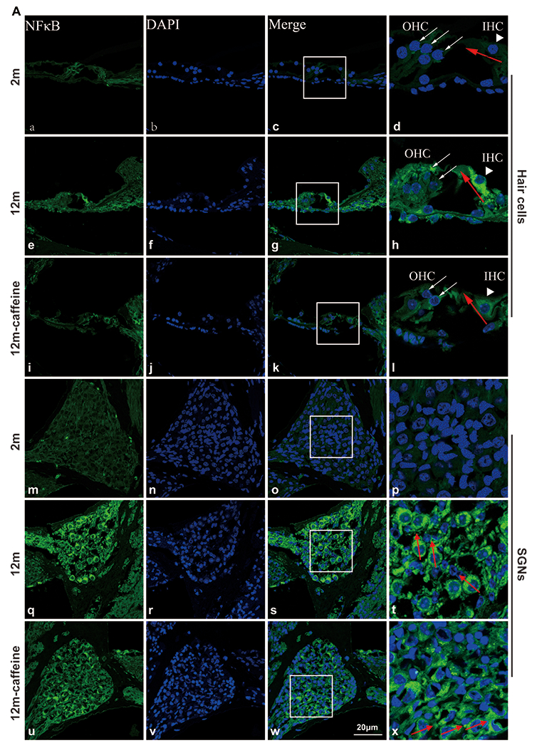
Methods: Caffeine was administered in drinking water to C57BL/6J mice from the age of 3 months to 12 months. The body weight, food intake and water intake of the mice were monitored during the experiment. The metabolic indicators of serum were detected by ELISA. The function of the hearing system was evaluated by ABR and hematoxylin and eosin staining of the cochlea. Genes' expression were detected by Q-PCR, immunofluorescencee and Western blot.
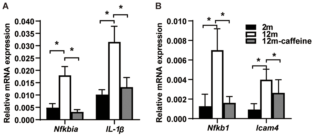
Results: The results showed that the ARHL mice exhibited impaired hearing and cochlear tissue compared with the young mice. However, the caffeine-treated ARHL mice showed improved hearing and cochlear tissue morphology. The expression of inflammation-related genes, such as TLR4, Myd88, NF-κB, and IL-1β, was significantly increased in the cochleae of ARHL mice compared with young mice but was down-regulated in the caffeine-treated cochleae.
Conclusions: Inflammation is involved in ARHL of mice, and long-term caffeine supplementation could ameliorate ARHL through the down-regulation of the TLR4/NF-κB inflammation pathway. Our findings provide a new idea for preventing ARHL and suggest new drug targets for ARHL treatment.
Copyright © 2024 The Author(s). Published by Wolters Kluwer Health, Inc. on behalf of Otology & Neurotology, Inc.
Conflict of interest statement
The authors disclose no conflicts of interest.
Figures


Similar articles
-
Mahonia bealei (Fort.) Carr. Leaf extract modulates the TLR2/MyD88/NF-κB signaling pathway to inhibit PGN-induced inflammation in RAW264.7 cells.J Ethnopharmacol. 2025 Mar 26;344:119510. doi: 10.1016/j.jep.2025.119510. Epub 2025 Feb 17. J Ethnopharmacol. 2025. PMID: 39971016
-
Identifying a gene signature for age-related hearing loss through machine learning and revealing the effect of the CTSS on the mice cochlea.Biogerontology. 2025 Jun 3;26(3):119. doi: 10.1007/s10522-025-10261-8. Biogerontology. 2025. PMID: 40461927
-
Whole-Genome DNA Methylation Analysis in Age-Related Hearing Loss.Genes (Basel). 2025 Apr 29;16(5):526. doi: 10.3390/genes16050526. Genes (Basel). 2025. PMID: 40428348 Free PMC article.
-
The Black Book of Psychotropic Dosing and Monitoring.Psychopharmacol Bull. 2024 Jul 8;54(3):8-59. Psychopharmacol Bull. 2024. PMID: 38993656 Free PMC article. Review.
-
Interventions to prevent occupational noise-induced hearing loss.Cochrane Database Syst Rev. 2017 Jul 7;7(7):CD006396. doi: 10.1002/14651858.CD006396.pub4. Cochrane Database Syst Rev. 2017. PMID: 28685503 Free PMC article.
Cited by
-
Bioinformatics approach reveals the critical role of inflammation-related genes in age-related hearing loss.Sci Rep. 2025 Jan 21;15(1):2687. doi: 10.1038/s41598-024-83428-x. Sci Rep. 2025. PMID: 39837906 Free PMC article.
-
Investigating the causal relationship between inflammation and multiple types of hearing loss: a multi-omics approach combining Mendelian randomization and molecular docking.Front Neurol. 2024 Nov 28;15:1422241. doi: 10.3389/fneur.2024.1422241. eCollection 2024. Front Neurol. 2024. PMID: 39677857 Free PMC article.
References
-
- Slade K, Plack CJ, Nuttall HE. The Effects of Age-Related Hearing Loss on the Brain and Cognitive Function. Trends Neurosci. 2020;43(10):810–21. - PubMed
-
- Franceschi C, Campisi J. Chronic Inflammation (Inflammaging) and Its Potential Contribution to Age-Associated Diseases. Journals of Gerontology. 2014;69(Suppl 1):S4–S9. - PubMed
-
- Hayden MS, Ghosh S. Shared principles in NF-kappaB signaling. Cell. 2008;132(3):344–62. - PubMed
MeSH terms
Substances
Grants and funding
LinkOut - more resources
Full Text Sources
