Review
. 2024 Feb 14;291(2016):20231304.
doi: 10.1098/rspb.2023.1304. Epub 2024 Feb 7.

Unpacking the navigation toolbox: insights from comparative cognition

Affiliations
Review

Unpacking the navigation toolbox: insights from comparative cognition

Kate J Jeffery et al. Proc Biol Sci. .

Abstract

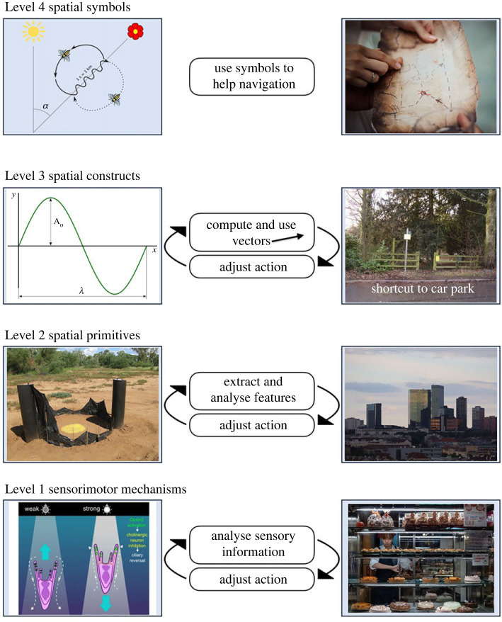
The study of navigation is informed by ethological data from many species, laboratory investigation at behavioural and neurobiological levels, and computational modelling. However, the data are often species-specific, making it challenging to develop general models of how biology supports behaviour. Wiener et al. outlined a framework for organizing the results across taxa, called the 'navigation toolbox' (Wiener et al. In Animal thinking: contemporary issues in comparative cognition (eds R Menzel, J Fischer), pp. 51-76). This framework proposes that spatial cognition is a hierarchical process in which sensory inputs at the lowest level are successively combined into ever-more complex representations, culminating in a metric or quasi-metric internal model of the world (cognitive map). Some animals, notably humans, also use symbolic representations to produce an external representation, such as a verbal description, signpost or map that allows communication of spatial information or instructions between individuals. Recently, new discoveries have extended our understanding of how spatial representations are constructed, highlighting that the hierarchical relationships are bidirectional, with higher levels feeding back to influence lower levels. In the light of these new developments, we revisit the navigation toolbox, elaborate it and incorporate new findings. The toolbox provides a common framework within which the results from different taxa can be described and compared, yielding a more detailed, mechanistic and generalized understanding of navigation.

Keywords: cognitive map; navigation; route; spatial cognition; vector; wayfinding.


Conflict of interest statement

We declare we have no competing interests.

Figures

Figure 1.
Figure 1.
The four levels of spatial representation. The left column shows illustrative cases with non-human animals and the right with humans. At Level 1, sensory information is used directly to guide the actions used in navigation. Left: a sea urchin larva swims upwards until the light level crosses a brightness threshold, whereupon it reverses (from [4]). Right: in a strange city, humans might ‘follow their nose’ to a bakery. Level 2: simple spatial features are extracted from sensory information. Example shows the use of views to guide navigation. Left: an ant at the start of a journey home using an artificial panoramic skyline for guidance that mimics the actual skyline [5]. Right: in open space, humans might also use the broad panorama for guidance. Level 3: spatial primitives of direction and distance are combined as vectors to guide navigation. Left: Drosophila flies may code and transform vectors in the form of spatial sine waves [6]. Right: Humans can learn to compute shortcuts to non-distinct locations, travelling in a particular direction for an approximate distance to reach their destination. Level 4: symbols constructed or communicated by others help in navigation. Left: the renowned waggle dance of the honeybee. Right: humans often use maps to help navigation. Figure credits. Level 1, photophobia in sea urchin: from [4], the open-source publication (licence: https://creativecommons.org/licenses/by/4.0/). Level 1, bakery: from Wikimedia creative commons (licence: https://creativecommons.org/licenses/by-sa/4.0/deed.en), author: Reinhold Möller. Level 2, skyline for ants: photo by Paul Graham. Level 2, city skyline: from Wikimedia creative commons (licence: https://creativecommons.org/licenses/by/4.0/deed.en), author: Marte007. Level 3, sine wave: from Wikimedia creative commons (licence: https://creativecommons.org/licenses/by-sa/3.0/deed.en), author: badseed, using work by Josemontero9 and José Luis Gálvez. Level 3, shortcut: from Wikimedia creative commons (licence: https://creativecommons.org/licenses/by-sa/2.0/deed.en), author: Anthony Vosper. Level 4, waggle dance: from Wikimedia creative commons (licence: https://creativecommons.org/licenses/by-sa/2.5/deed.en), author not named. Level 4, treasure map: from Pixabay (licence: https://pixabay.com/service/license-summary/), author: Pexels.
Figure 2.
Figure 2.
Sensorimotor processes allow simple forms of navigation towards or away from stimuli, such as in chemotaxis or beaconing. (a) Chemotaxis in algae in response to the pheromone lamoxirene [11]. (b,c) Beacons, positive, encouraging approach (b), and negative, encouraging avoidance (c). Figure credits: (b) image from https://commons.wikimedia.org/wiki/File:Eiffel_Tower_-_Paris_-_2016.JPG by Brian Lee is licensed under the Creative Commons Attribution-Share Alike 4.0 International license. (c) Image from https://commons.wikimedia.org/wiki/File:Mevagissey_lighthouse_(9453).jpg by Nilfanion is licensed under the Creative Commons Attribution-Share Alike 4.0 International license.
Figure 3.
Figure 3.
The neural basis of the spatial primitive of direction, recorded in two different taxa: rodents and insects. (a) Left: neurons from one of the head direction (HD) cell regions are recorded as a rat explores a cylindrical chamber in which direction is indicated by a single landmark (white card on the wall). Right: polar plot of the firing rate of a single HD neuron is plotted as a function of the facing direction of the animal. Accumulated time spent facing each of the possible directions is shown in grey. This neuron fired maximally (green line) when the rat faced ‘southeast’ (blue plot). (b) Neuronal activity recorded by a scanning microscope from a fruit fly as it ‘walked’ on an air-cushioned ball that controlled a video display simulating a real environment. Right (top): plots showing successive points in time as a visual cue (pale blue bar) moved around the screen, coupled to the actions of the fly. Right (bottom): heat plots showing hot-spots of neural activity in the circular region of the fly brain that maintain a consistent relationship to the visual cue. The same effect was seen in darkness (not shown), indicating that this is not just a visual response, but one that integrates the sensorimotor tools of vision and locomotion. Figure adapted from fig. 1 of [26]; published with permission (please note the rights are held by a third party).
Figure 4.
Figure 4.
Activity of rodent grid cells. (a) Schematic showing the recording of a single entorhinal grid cell. A rat explores a square arena, with its path (grey line) tracked by an overhead camera. Inset oscilloscope trace shows neuronal spikes (action potentials; highlighted with red squares) from a single neuron. The red squares on the arena depict those same spikes placed at the location where the rat was when they were emitted. They congregate in restricted regions of the arena. (b) When the spikes plotted as in (a) are accumulated over a trial they form regularly spaced firing fields, indicative of odometry (distance-tracking). For simple symmetrical environments like this square platform, the firing fields form rows with a stable orientation. (c) The regular spacing indicates the integration of both distance and direction. (d) In three dimensions, this regularity breaks down, although the discreteness of the firing fields remains [41].
Figure 5.
Figure 5.
Two forms of border-related firing: egocentric (a) and allocentric (b). The plots show spikes for a single neuron overlaid on the path of a rat in grey, as previously. The egocentric boundary cell in (a) was recorded in the retrosplenial cortex [55]. It is ‘egocentric’ because the cell fires when the border has a given directional relationship to the animal, as shown by the colour coding. The boundary vector cell in (b) only fired when the rat was against a boundary lying at a given allocentric direction (e.g. north).
Figure 6.
Figure 6.
Place cells. (a) Data from a rat exploring two connected boxes as shown in figure 4 but with the path of the rat in yellow for one box and brown for the other. The red dots are spikes from each of the three simultaneously recorded neurons. Note that the cells fire differently in the two boxes, and also that their firing locations overlap considerably. (b) Place cells also encode sequences of locations. In the schematic, a rat is either walking (left) or resting/sleeping (right). The spikes from two cells are shown, aligned along the path of the rat and also across time. Left: during walking, the spikes occur sequentially in both time and place. Right: during resting, the same temporal sequence of spikes spontaneously recurs, suggesting reactivation of ‘memory’ for the route.

Similar articles

Cited by

References

    1. Gladwin T. 2009. East is a big bird: navigation and logic on Puluwat Atoll. Cambridge, MA: Harvard University Press.
    1. Krakauer JW, Ghazanfar AA, Gomez-Marin A, MacIver MA, Poeppel D. 2017. Neuroscience needs behavior: correcting a reductionist bias. Neuron 93, 480-490. (10.1016/j.neuron.2016.12.041) - DOI - PubMed
    1. Wiener J, et al. 2011. Animal navigation: a synthesis. In Animal thinking: contemporary issues in comparative cognition (eds R Menzel, J Fischer), pp. 51–76. Cambridge, MA: MIT Press. (10.7551/mitpress/9780262016636.003.0005) - DOI
    1. Brodrick E, Jékely G. 2023. Photobehaviours guided by simple photoreceptor systems. Anim. Cogn. 26, 1817-1835. (10.1007/s10071-023-01818-6) - DOI - PMC - PubMed
    1. Graham P, Cheng K. 2009. Ants use the panoramic skyline as a visual cue during navigation. Curr. Biol. 19, R935-R937. (10.1016/j.cub.2009.08.015) - DOI - PubMed

LinkOut - more resources