. 2024 Feb 6;18(7):5684-5697.
doi: 10.1021/acsnano.3c11579. Online ahead of print.

Colloidal Aziridinium Lead Bromide Quantum Dots

Affiliations

Colloidal Aziridinium Lead Bromide Quantum Dots

Maryna I Bodnarchuk et al. ACS Nano. .

Abstract

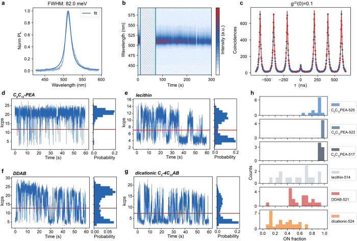
The compositional engineering of lead-halide perovskite nanocrystals (NCs) via the A-site cation represents a lever to fine-tune their structural and electronic properties. However, the presently available chemical space remains minimal since, thus far, only three A-site cations have been reported to favor the formation of stable lead-halide perovskite NCs, i.e., Cs+, formamidinium (FA), and methylammonium (MA). Inspired by recent reports on bulk single crystals with aziridinium (AZ) as the A-site cation, we present a facile colloidal synthesis of AZPbBr3 NCs with a narrow size distribution and size tunability down to 4 nm, producing quantum dots (QDs) in the regime of strong quantum confinement. NMR and Raman spectroscopies confirm the stabilization of the AZ cations in the locally distorted cubic structure. AZPbBr3 QDs exhibit bright photoluminescence with quantum efficiencies of up to 80%. Stabilized with cationic and zwitterionic capping ligands, single AZPbBr3 QDs exhibit stable single-photon emission, which is another essential attribute of QDs. In particular, didodecyldimethylammonium bromide and 2-octyldodecyl-phosphoethanolamine ligands afford AZPbBr3 QDs with high spectral stability at both room and cryogenic temperatures, reduced blinking with a characteristic ON fraction larger than 85%, and high single-photon purity (g(2)(0) = 0.1), all comparable to the best-reported values for MAPbBr3 and FAPbBr3 QDs of the same size.

Keywords: aziridinium; ligands; nanocrystals; perovskite; photoluminescence; quantum dots.


Conflict of interest statement

The authors declare no competing financial interest.

Figures

Figure 1
Figure 1
(a) Calculated Goldschmidt tolerance factors for different cations (cesium, methylammonium, aziridinium, and formamidinium) in the APbBr3 perovskite lattice. (b) Top panel: a reaction scheme; bottom panel: an overview of carboxylic and phosphonic acids as well as ligands used in the synthesis. (c) Optical absorption and (d) PL spectra of purified AZPbBr3 NCs ranging from 4.5 to 14 nm in size (for visualization purposes, a cumulative vertical offset was applied to each subsequent spectra). (e) A high-angle annular dark field-scanning transmission electron microscopy (HAADF-STEM) image of purified 8 nm NCs with a high-resolution HAADF-STEM image of few single NC in the inset. (f) Size-dependent (absorption) band gap in AZPbBr3 NCs for sizes obtained via TEM (green) and SAXS (gray); the experimental data sets (open squares, with error bars denoting the standard deviation) were fitted by a semiempirical sizing curve (solid line), with the bulk band gap (dashed lines) as one of the fit parameters.
Figure 2
Figure 2
(a) The fit of experimental SAXS data from an AZPbBr3 NC colloidal suspension (gray line) via an analytical model (black line) yields cuboids with edge lengths of 8.27 ± 0.12, 8.28 ± 0.93, and 8.60 ± 0.48 nm, respectively. (b) Solvent-subtracted synchrotron WAXTS data (black line) and the best fit (green line) of AZPbBr3 NCs using the split cubic model with locally tilted/disordered PbBr6 octahedra (for a more extended discussion, see the Supporting Information); the inset shows a 2D map of the refined (number-based) log-normal size distribution function (Dab: the diameter of the circle of area equivalent to the prisms basal plane and Lc, the height of the prismatic clusters). (c) The split cubic structural model of AZPbBr3 NCs (the four Br of each PbBr6 octahedra, shown in green, have 1/4 site occupancy factor each, i.e., only one out of four is stochastically present in each site) and the model of AZ cation formed by disordering in 12 symmetry-equivalent orientations (4 equivalent geometrical orientations × 3 “elemental” C–C/N dispositions, H atoms were omitted, similar to bulk AZPbBr3). (d) The temperature dependence of the tetragonal unit cell parameters a′ = a/√2 and c for a weakly distorted (ca. 1%) cubic lattice, plotted in the entire 11–290 K range, showing an anomaly below 90 K. (e) The temperature dependence of the fwhm0 parameter (the θ-dependence of the peak width is described according to the relation fwhm(θ) = fwhm0/cos θ), which suggests additional peak broadening (hidden splitting) below 90 K.
Figure 3
Figure 3
(a) Solution 1H NMR spectra of C8C12–PEA (black line) and DDAB-capped (blue line) AZPbBr3 NCs in cyclohexane-d12. (b) Solution 31P NMR spectra of C8C12–PEA-capped AZPbBr3 NCs synthesized without (top) and with (middle) addition of alkylphosphonic acid (for instance, octylphosphonic acid-C8PAc) during the synthesis and after their further digestion with DMSO-d6 (bottom). (c) Solid-state 207Pb NMR spectrum of C8C12–PEA-capped AZPbBr3 NCs.
Figure 4
Figure 4
Room-temperature PL of single AZPbBr3 QDs with various surface capping ligands. (a) PL spectrum of a single C8C12–PEA-capped AZPbBr3 QD displaying narrow-band emission with a fwhm of 82 meV. (b) Spectra series of a C8C12–PEA-capped QD exhibiting spectrally stable PL and small intensity variations. The highlighted time period (shaded area without PL spectral detection) corresponds to the acquisition of the second-order correlation function (g(2)(t), shown in panel (c)) and blinking trace by a Hanbury–Brown and Twiss experiment. (c) Second-order photon–photon correlation of a C8C12–PEA-capped AZPbBr3 QD displaying high single-photon purity (g(2)(0) = 0.1). (d–g) Representative PL blinking traces (10 ms binning time) of a QD capped with branched C8C12–PEA ligands (panel (d)), lecithin (panel (e)), DDAB (panel (f)), and dicationic amine C3–4C12AB (panel (g)). (h) Histograms of the fraction of time that single QDs spend in their bright (ON) state. The numbers after the ligand name indicate the ensemble PL central wavelength of the respective sample.
Figure 5
Figure 5
Exciton fine-structure of single AZPbBr3 QDs at 4 K. (a) Time series of a single QD (1 s integration time and 1800 g/mm grating); sub-meV spectral diffusion allows to resolve the exciton fine structure. This QD exhibits a doublet fine structure with peaks denoted as FS1 and FS2. The inset shows an associated histogram of the peak energies (bars), fitted with a Gaussian function (lines). (b) The PL spectra of one single QD measured at two different angles of a linear polarizer in the detection path: 45° (blue) and 135° (gray); this QD has a doublet exciton fine structure. The inset shows a polar plot with the respective PL intensities, as a function of the polarizer angle; both doublet sublevels exhibit highly linear polarization, oriented perpendicular to each other; (c, d) Fine-structure splitting energy (FSS) for all of the single QDs (capped with DDAB or C8C12–PEA ligands) exhibit doublet sublevels (panel (c)) or triplet sublevels (panel (d)). The insets in panels (c) and (d) show representative spectra for doublet and triplet exciton fine structures, respectively. (e) Second-order correlation function g(2)(τ) of a single AZPbBr3 QD under 0.3 μJ/cm2 before (dark gray line) and after (blue line) spectrally filtering out the biexciton emission by a tunable short-pass filter.

Similar articles

Cited by

References

    1. Dey A.; Ye J.; De A.; Debroye E.; Ha S. K.; Bladt E.; Kshirsagar A. S.; Wang Z.; Yin J.; Wang Y.; et al. State of the Art and Prospects for Halide Perovskite Nanocrystals. ACS Nano 2021, 15, 10775–10981. 10.1021/acsnano.0c08903. - DOI - PMC - PubMed
    1. Akkerman Q. A.; Rainò G.; Kovalenko M. V.; Manna L. Genesis, Challenges and Opportunities for Colloidal Lead Halide Perovskite Nanocrystals. Nat. Mater. 2018, 17, 394–405. 10.1038/s41563-018-0018-4. - DOI - PubMed
    1. Kovalenko M. V.; Protesescu L.; Bodnarchuk M. I. Properties and Potential optoelectronic Applications of Lead Halide Perovskite Nanocrystals. Science 2017, 358, 745–750. 10.1126/science.aam7093. - DOI - PubMed
    1. Protesescu L.; Yakunin S.; Bodnarchuk M. I.; Krieg F.; Caputo R.; Hendon C. H.; Yang R. X.; Walsh A.; Kovalenko M. V. Nanocrystals of Cesium Lead Halide Perovskites (CsPbX3, X = Cl, Br, and I): Novel Optoelectronic Materials Showing Bright Emission with Wide Color Gamut. Nano Lett. 2015, 15, 3692–3696. 10.1021/nl5048779. - DOI - PMC - PubMed
    1. Rainò G.; Nedelcu G.; Protesescu L.; Bodnarchuk M. I.; Kovalenko M. V.; Mahrt R. F.; Stöferle T. Single Cesium Lead Halide Perovskite Nanocrystals at Low Temperature: Fast Single-Photon Emission, Reduced Blinking, and Exciton Fine Structure. ACS Nano 2016, 10, 2485–2490. 10.1021/acsnano.5b07328. - DOI - PMC - PubMed