. 2024 Dec;42(12):1822-1830.
doi: 10.1038/s41587-023-02106-x. Epub 2024 Feb 6.

Efficient prime editing in two-cell mouse embryos using PEmbryo

Affiliations

Efficient prime editing in two-cell mouse embryos using PEmbryo

Rebecca P Kim-Yip et al. Nat Biotechnol. 2024 Dec.

Abstract

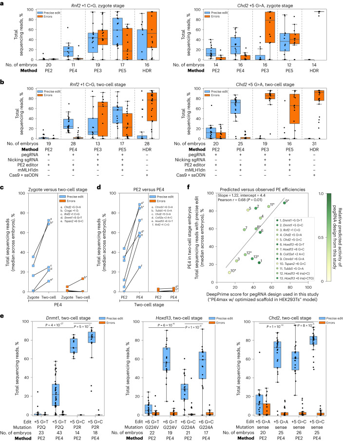
Using transient inhibition of DNA mismatch repair during a permissive stage of development, we demonstrate highly efficient prime editing of mouse embryos with few unwanted, local byproducts (average 58% precise edit frequency, 0.5% on-target error frequency across 13 substitution edits at 8 sites), enabling same-generation phenotyping of founders. Whole-genome sequencing reveals that mismatch repair inhibition increases off-target indels at low-complexity regions in the genome without any obvious phenotype in mice.


Conflict of interest statement

Competing interests: B.A. is an advisory board member with options for Arbor Biotechnologies and Tessera Therapeutics. B.A. holds equity in Celsius Therapeutics. D.R.L. is a consultant and equity holder of Beam Therapeutics, Prime Medicine, Pairwise Plants, Chroma Medicine, Resonance Medicine, Exo Therapeutics and Nvelop Therapeutics, companies that use genome editing or engineering. P.J.C. is currently an employee of Prime Medicine. J.E.T. is an advisor to Nereid Therapeutics and Prolific Machines. E.A.I. has pending interest in VUS Diagnostics. B.A., D.R.L. and P.J.C. are co-inventors on prime editing patent applications filed through their respective institutions. The other authors declare no competing interests.

Figures

Fig. 1
Fig. 1. Dominant negative mMLH1 and delivery at the two-cell stage improves prime editing in mouse embryos.
a, Percentages of total reads containing only the indicated precise edit (blue) or errors (orange) in Rnf2 (left) or in Chd2 (right). Each data point represents an individual embryo edited at the zygote stage. Editing conditions indicated in b. b, Same as a, except each data point represents an individual embryo edited at the two-cell stage. HDR, homology-directed repair; ssODN, single-strand oligonucleotide donor. c, Median precise edit (blue) and error (orange) frequencies across embryos microinjected with PE4 components (editor mRNA, pegRNA, mMLH1dn mRNA) at the zygote or two-cell stage. Plot includes data also represented in a, b and e. d, Same as c, except plot represents data from embryos microinjected at the two-cell stage with PE2 components (editor mRNA, pegRNA) or PE4 components (editor mRNA, pegRNA, mMLH1dn mRNA). Plot includes data also represented in b and e. e, Percentages of total reads containing only the indicated precise edit (blue) or errors (orange) from individual embryos microinjected at the two-cell stage. Plots include Chd2 results from b. f, Comparison of predicted prime editing efficiencies (DeepPrime score) from a deep-learning-based model trained on editing results in HEK293T cells using an optimized prime editor (PEmax) and hMLH1dn to observed prime editing (PE) efficiencies in mouse embryos microinjected at the two-cell stage with PE4 components (editor mRNA, pegRNA, mMLH1dn mRNA) from this study. Each dot represents the specific pegRNA design used in our study. Color shade indicates the relative predicted score of the pegRNA compared to the maximum score predicted by the DeepPrime-FT model across all feasible pegRNA designs (Methods) for a given target site/edit. Pearson correlation coefficient (r = 0.68) reported with P value (P = 0.01) from two-sided t-test. Dashed line represents fit from linear least-squares regression. Throughout our study, asterisks specify use of the optimized PEmax editor (PE2*, PE4* methods), as opposed to the PE2 editor (PE2, PE4 methods). Data in af are compiled from multiple experiments (Supplementary Tables 7–9 and Methods). For c and d, lowercase letters indicate edit and black lines connect results for the same edit across conditions. For ce, P values are from two-sided Student’s t-tests. For box plots, boxes indicate the median and interquartile range (IQR) for each group of embryos with whiskers extending 1.5 × IQR past the upper and lower quartiles.
Fig. 2
Fig. 2. WGS after transient MMR inhibition in embryos.
a, Percentages of total reads containing the precise +5 G > A edit (blue) or errors (orange) in Chd2 from ear clips of 2- to 3-week-old mice developed from embryos microinjected with PE4 components (PE2 editor mRNA, pegRNA, mMLH1dn mRNA) at the two-cell stage. Data compiled from multiple experiments (Supplementary Table 13 and Methods). b, Pedigree of ‘PE4 family’ (top). Black indicates the ‘treated’ group of select progeny microinjected at the two-cell stage with PE4 components (PE2 editor mRNA, Chd2 + 5 G > A pegRNA, mMLH1dn mRNA). Unshaded family members indicate mice/embryos treated as the ‘control’ group, including sibling progeny microinjected at the two-cell stage with PE2 editor mRNA only. Percentages indicate precise edit efficiency at E12.5 as determined by WGS. Plot (bottom) compares editing frequencies in treated embryos across sequencing methods (target versus whole-genome sequencing). Superscripts denote individual progeny from ‘PE4 family’. Dashed line represents x = y. c, Total unique SNVs (left) and total unique indels (right) detected in members of the ‘PE4 family’ after joint genotyping (black line indicates mean from each group, P values from two-sided Welch’s t-tests). d, Cumulative frequencies of unique SNVs (left) or unique indels (right) by type for members of the ‘PE4 family’. F, female; M, male. e, Fraction of unique −1 bp deletions directly adjacent to poly(A/T) nucleotide tracts in treated and control mice/embryos from each indicated family (P values from two-sided Welch’s t-test). Treated and control groups are defined in b and f. f, Pedigrees of additional mouse families. Black denotes treated groups. Unshaded siblings comprise control groups. For the ‘PE2* family’ (left), treated embryos were microinjected with PE2* components (PEmax mRNA, Chd2 + 5 G > A pegRNA) at the two-cell stage, whereas control embryos were microinjected with pegRNA only. For the ‘mMLH1dn family’ (right), treated embryos were microinjected with mMLH1dn mRNA and the Chd2 + 5 G > A pegRNA (but no editor) at the two-cell stage, whereas control embryos were microinjected with pegRNA only. One control embryo (not indicated) was omitted from analysis for this family after sequencing failed quality control (Methods). Percentages indicate precise edit efficiency in treated embryos at E12.5. g, Number of unique indels detected in treated embryos in each family (n = 3, PE4 family; n = 2, PE2* family; n = 3, mMLH1dn family) relative to the average of control mice/embryos from the same family. Data points represent fold-change for individual mice/embryos. Bars indicate the mean difference. For pedigree diagrams, red dashed boxes indicate mice/embryos subjected to WGS. For box plots, boxes indicate the median and IQR of each group with whiskers extending 1.5 × IQR (a) and 2 × IQR (e) past the upper and lower quartiles.
Fig. 3
Fig. 3. Prime editing with PEmbryo allows for same-generation phenotyping of a substitution edit in mice.
a, Percentages of total reads containing the precise +6 G > T edit (blue) or errors (orange) in Hoxd13 from ear clips of 34 pups developed from embryos microinjected with PE4 components (PE2 editor mRNA, pegRNA and mMLH1dn mRNA) at the two-cell stage. Plot on the right depicts the same data as shown on the left, but with mice sorted into three groups based on the severity of the brachydactyly phenotype of the fifth digit on the front limbs (none, moderate or severe). Boxes indicate the median and IQR of each group with whiskers extending 1.5 × IQR past the upper and lower quartiles. b, Schematic for breeding of N0 founder mice to generate N1 and N1F1 generations with different genotypes for Hoxd13 + 6 G > T (G224V). Checkered texture indicates mosaic pattern of edits in the founder mouse. Light blue shading represents heterozygous mice with one copy of the edit. Dark blue shading represents homozygous mice with two copies of the edit. Percentages of the N1F1 generation indicate the expected (not actual) mendelian frequencies of each genotype. c, Representative images of left and right front paws of pups from Hoxd13-edited N0 founder mice sorted by phenotype severity before sequencing. Control images from comparably aged (18-day-old), Chd2-edited pups from the same microinjection experiment. Asterisks indicate fifth digits. d, Representative images of left and right front paws of pups from N1 mice heterozygous for Hoxd13 + 6 G > T (G224V). e, Representative images of left and right front paws of pups from N1F1 mice sorted by genotype: wild-type, heterozygous or homozygous for Hoxd13 + 6 G > T (G224V). f, Sanger sequencing traces for N1F1 mice wild-type, heterozygous or homozygous for Hoxd13 + 6 G > T (G224V). Yellow shading indicates the target site. Trace colors correspond to the base call at each site: T, thymine (red), C, cytosine (blue), A, adenine (green), G, guanine (black).

References

    1. Harms, D. et al. Mouse genome editing using the CRISPR/Cas system. Curr. Protoc. Hum. Genet.83, 15.7.1–15.7.27 (2014). - PMC - PubMed
    1. Clark, J. F., Dinsmore, C. J. & Soriano, P. A most formidable arsenal: genetic technologies for building a better mouse. Genes Dev.34, 1256–1286 (2020). - PMC - PubMed
    1. Modzelewski, A. J. et al. Efficient mouse genome engineering by CRISPR-EZ technology. Nat. Protoc.13, 1253–1274 (2018). - PMC - PubMed
    1. Wang, H. et al. One-step generation of mice carrying mutations in multiple genes by CRISPR/Cas-mediated genome engineering. Cell153, 910–918 (2013). - PMC - PubMed
    1. Wilde, J. J. et al. Efficient embryonic homozygous gene conversion via RAD51-enhanced interhomolog repair. Cell184, 3267–3280.e18 (2021). - PMC - PubMed

LinkOut - more resources