

Inflammation-related pathology in the olfactory epithelium: its impact on the olfactory system in psychotic disorders
- PMID: 38321120
- PMCID: PMC11189720
- DOI: 10.1038/s41380-024-02425-8
Inflammation-related pathology in the olfactory epithelium: its impact on the olfactory system in psychotic disorders
Abstract
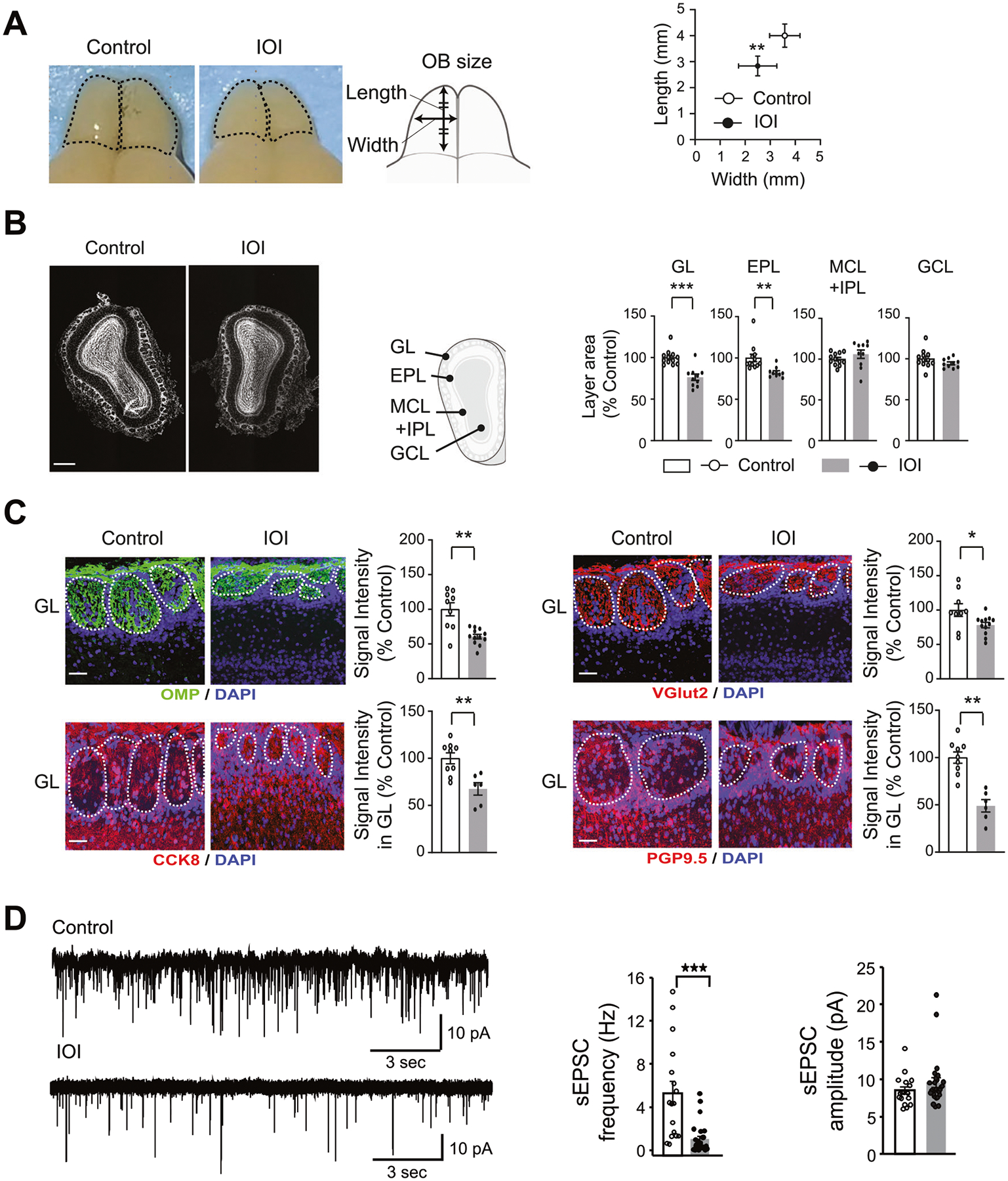
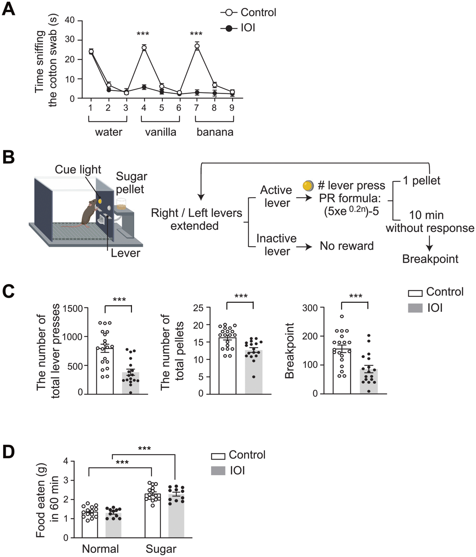
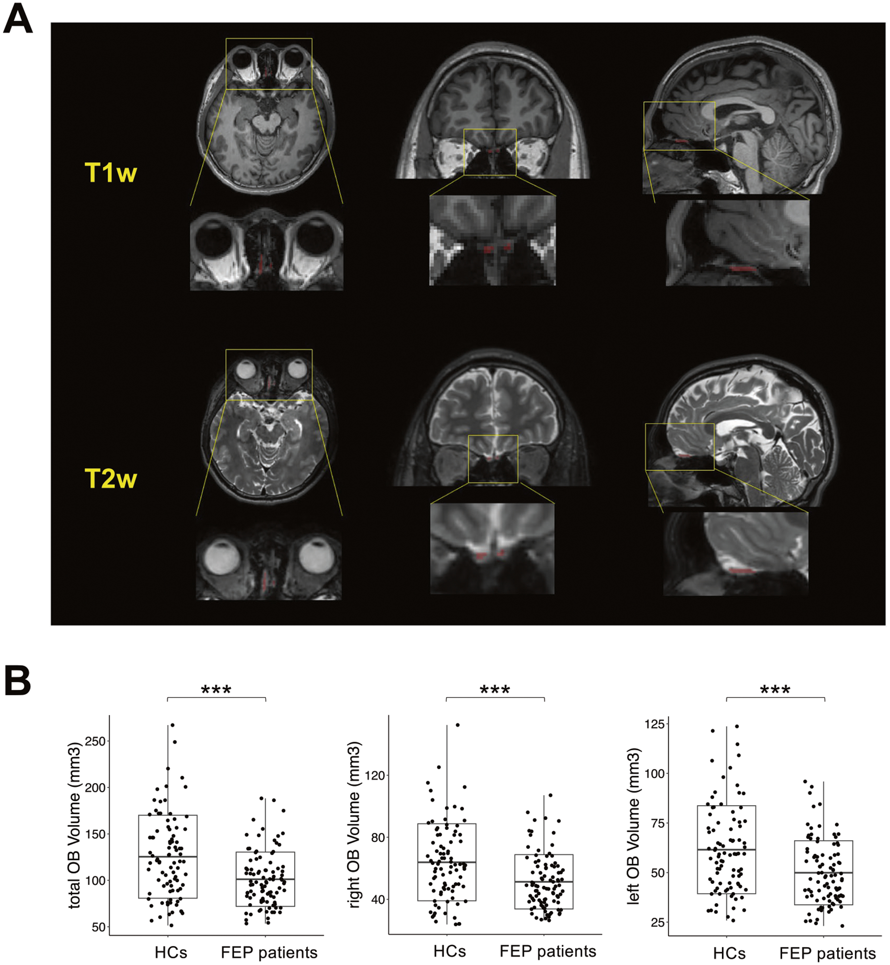
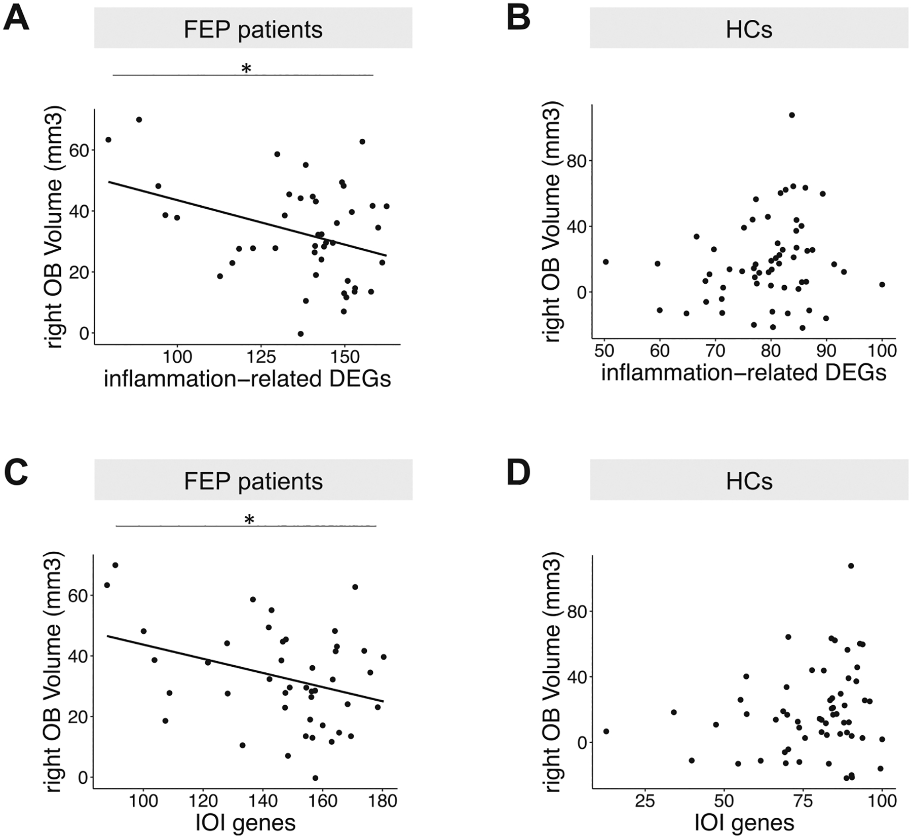
Smell deficits and neurobiological changes in the olfactory bulb (OB) and olfactory epithelium (OE) have been observed in schizophrenia and related disorders. The OE is the most peripheral olfactory system located outside the cranium, and is connected with the brain via direct neuronal projections to the OB. Nevertheless, it is unknown whether and how a disturbance of the OE affects the OB in schizophrenia and related disorders. Addressing this gap would be the first step in studying the impact of OE pathology in the disease pathophysiology in the brain. In this cross-species study, we observed that chronic, local OE inflammation with a set of upregulated genes in an inducible olfactory inflammation (IOI) mouse model led to a volume reduction, layer structure changes, and alterations of neuron functionality in the OB. Furthermore, IOI model also displayed behavioral deficits relevant to negative symptoms (avolition) in parallel to smell deficits. In first episode psychosis (FEP) patients, we observed a significant alteration in immune/inflammation-related molecular signatures in olfactory neuronal cells (ONCs) enriched from biopsied OE and a significant reduction in the OB volume, compared with those of healthy controls (HC). The increased expression of immune/inflammation-related molecules in ONCs was significantly correlated to the OB volume reduction in FEP patients, but no correlation was found in HCs. Moreover, the increased expression of human orthologues of the IOI genes in ONCs was significantly correlated with the OB volume reduction in FEP, but not in HCs. Together, our study implies a potential mechanism of the OE-OB pathology in patients with psychotic disorders (schizophrenia and related disorders). We hope that this mechanism may have a cross-disease implication, including COVID-19-elicited mental conditions that include smell deficits.
© 2024. The Author(s), under exclusive licence to Springer Nature Limited.
Conflict of interest statement
COMPETING INTERESTS
The authors declare no competing interests.
Figures
Update of
-
Inflammation-related pathology in the olfactory epithelium: its impact on the olfactory system in psychotic disorders.bioRxiv [Preprint]. 2024 Jan 10:2022.09.23.509224. doi: 10.1101/2022.09.23.509224. bioRxiv. 2024. Update in: Mol Psychiatry. 2024 May;29(5):1453-1464. doi: 10.1038/s41380-024-02425-8. PMID: 36203543 Free PMC article. Updated. Preprint.
References
-
- Cohen AS, Brown LA, Auster TL. Olfaction, ‘olfiction,’ and the schizophrenia-spectrum: an updated meta-analysis on identification and acuity. Schizophr Res. 2012;135:152–7. - PubMed
-
- Kopala LC, Good K, Honer WG. Olfactory identification ability in pre- and post-menopausal women with schizophrenia. Biol Psychiatry. 1995;38:57–63. - PubMed
MeSH terms
Grants and funding
- R21 MH128765/MH/NIMH NIH HHS/United States
- R01 AG064093/AG/NIA NIH HHS/United States
- R01 NS108452/NS/NINDS NIH HHS/United States
- R55 DC006213/DC/NIDCD NIH HHS/United States
- R01 MH105660/MH/NIMH NIH HHS/United States
- R01 AI132590/AI/NIAID NIH HHS/United States
- MH-107730/U.S. Department of Health & Human Services | NIH | National Institute of Mental Health (NIMH)
- R01 MH107730/MH/NIMH NIH HHS/United States
- DC-006213/U.S. Department of Health & Human Services | NIH | National Institute on Deafness and Other Communication Disorders (NIDCD)
- R01 AG065168/AG/NIA NIH HHS/United States
- R01 DC020841/DC/NIDCD NIH HHS/United States
- R01 MH092443/MH/NIMH NIH HHS/United States
- MH-094268/U.S. Department of Health & Human Services | NIH | National Institute of Mental Health (NIMH)
- NS-108452/U.S. Department of Health & Human Services | NIH | National Institute of Neurological Disorders and Stroke (NINDS)
- DA-041208/U.S. Department of Health & Human Services | NIH | National Institute on Drug Abuse (NIDA)
- AG-064093/U.S. Department of Health & Human Services | NIH | National Institute on Aging (U.S. National Institute on Aging)
- P50 MH094268/MH/NIMH NIH HHS/United States
- MH-105660/U.S. Department of Health & Human Services | NIH | National Institute of Mental Health (NIMH)
- DC-016106/U.S. Department of Health & Human Services | NIH | National Institute on Deafness and Other Communication Disorders (NIDCD)
- AG-065168/U.S. Department of Health & Human Services | NIH | National Institute on Aging (U.S. National Institute on Aging)
- MH-092443/U.S. Department of Health & Human Services | NIH | National Institute of Mental Health (NIMH)
- AI-132590/Division of Intramural Research, National Institute of Allergy and Infectious Diseases (Division of Intramural Research of the NIAID)
- R01 DC016106/DC/NIDCD NIH HHS/United States
- MH-128765/U.S. Department of Health & Human Services | NIH | National Institute of Mental Health (NIMH)
- R01 DC006213/DC/NIDCD NIH HHS/United States
- R01 DA041208/DA/NIDA NIH HHS/United States
LinkOut - more resources
Full Text Sources
Medical
Miscellaneous
