. 2024 Feb 6;14(1):3044.
doi: 10.1038/s41598-024-53126-9.

High-throughput cell optoporation system based on Au nanoparticle layers mediated by resonant irradiation for precise and controllable gene delivery

Affiliations

High-throughput cell optoporation system based on Au nanoparticle layers mediated by resonant irradiation for precise and controllable gene delivery

T E Pylaev et al. Sci Rep. .

Abstract

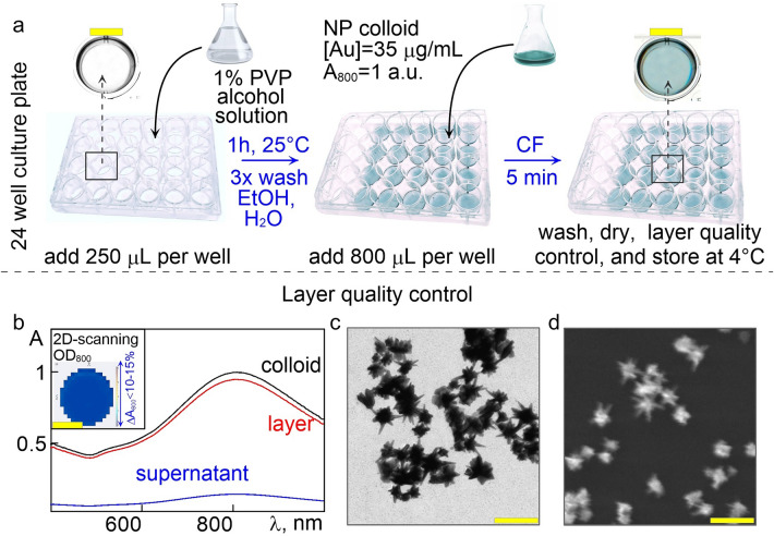
The development of approaches based on genetically modified cells is accompanied by a constant intensive search for new effective and safe delivery systems and the study of existing ones. Recently, we developed a new plasmonic nanoparticle layers-mediated optoporation system that can be proposed for precisely controlled, high-performance laser transfection compatible with broad types of cells and delivered objects of interest. The main goal of the present study is to demonstrate the broad possibilities and advantages of our system for optoporation of several mammalian cells, classified as "easy-to-transfect" cells, namely HeLa and CHO lines, and "hard-to-transfect" cells, namely A431 and RAW 264.7 cells. We show the efficient delivery of various sized cargo molecules: from small molecular dyes propidium iodide (PI) with molecular mass 700 Da, control plasmids (3-10 kb) to fluorophore-labeled dextranes with masses ranging from 10 kDa up to 100 kDa. The performance of optoporation was investigated for two types of laser sources, 800-nm continuous-wave laser, and 1064-nm ns pulsed laser. We provided a comparative study between our system and commercial agent Lipofectamine for transient transfection and stable transfection of HeLa cells with plasmids encoding fluorescent proteins. The quantitative data analysis using flow cytometry, Alamar blue viability assay, and direct fluorescence microscopy revealed higher optoporation efficacy for hard-to-transfect A431 cells and Raw 264.7 cells than lipofection efficacy. Finally, we demonstrated the optoporation performance at the single-cell level by successful delivering PI to the individual CHO cells with revealed high viability for at least 72 h post-irradiation.


Conflict of interest statement

The authors declare no competing interests

Figures

Figure 1
Figure 1
(a) Schematic representation of the AuNS layers preparation technology. (b) Normalized extinction spectra of the initial colloid, the supernatant after CF, and the obtained layer. The inset shows the spectrograms of the layer's uniformity. (c) Typical TEM image of as-prepared AuNS colloid. (d) SEM micrographs of AuNS layers. Scale bars correspond to 5 mm (enlarged scanned images of culture plate wells, a, b) and 100 nm (electron micrographs of particles, c, d).
Figure 2
Figure 2
Coordinate mesh gridding via AuNS layer laser engraving. (a) Scan image of the AuNS layer on the well bottom of the 24-well plate. (b) Schematic representation of laser engraving with set laser parameters. (c) Scan image of the grid-marked AuNS layer. (d) Phase-contrast micrograph of the gridded AuNS layer, 20 × objective.
Figure 3
Figure 3
The experimental setup of optoporation systems: with CW laser source (the upper row), and pulsed laser source (the below row). (a) Photoimages of the optoporation systems. (b) Photoimages of the 24-well plate with AuNS layers under the laser exposure. The red light from the satellite diode visualizes the irradiated well. (c) Schematic representation of the experimental setup and the working principle of the optoporation system. (d) Fluorescent microscopy images of optoporated HeLa cells with the delivered PI dye. Scale bars correspond to 100 µm.
Figure 4
Figure 4
(a) Relative number of sample and control HeLa cells expressing Gluc and GFP, determined by FACS 72 h after optotransfection with CW laser, pulsed laser and lipofection with LF. (b-d) Algorithm for post-processing of the FACS data. The inset microimages, acquired by AMNIS instrument on three emission channels under 488 nm excitation, depict the cell debris (b, 1–4) the cell aggregates (b), the control cell (c), and experimental cell (d) samples taken on three channels of the cytometer (1, 2, 3). The mean data is represented in the bars (a) with the standard deviations at a significance level of p ≤ 0.05 set for the error bars.
Figure 5
Figure 5
(a) Luciferase activity of the optotransfected HeLa-GLuc+ cells, lipofected cells, and control cells. (b) Representative CLSM microimages of optotransfected HeLa-mCherry + captured throughout the selection process until the complete monolayer with stably transfected cells was obtained; and after the cycle of cryopreservation/depreservation. The days of the experiment are designated below in arabic, the corresponding passages in roman numerals. The scale bars are 50 µm.
Figure 6
Figure 6
Optotransfection of "hard-to-transfect" A431 cells. (a-c) CLSM Microimages of the cells in a fluorescent and phase-contrast mode: (a) control cells, (b) optoporated cells by CW laser, and (c) lipofected cells. (d) Determination of the lower threshold starting cell monolayer confluence. (e) FACS data: the relative number of pDNA-positive cells optoporated by CW laser and pulsed laser, lipofected cells, normalized to control (intact cells). (f) Cell viability data recorded by Alamar blue assay. The scale bars correspond to 50 µm. The mean data is represented in the bars (e, f) with the standard deviations at a significance level of p ≤ 0.05 set for the error bars.
Figure 7
Figure 7
Optoporation of a single cell. (a-c) fluorescence microimages and (d-f) phase-contrast microimages of CHO cells with a magnification of the single optoporated cell (surrounded by a dotted-line circle); the yellow dotted line shows the trajectory of the pulsed laser for marking the area around the single optoporated cell. The scale bars correspond to 50 µm.

References

    1. Li H, et al. Applications of genome editing technology in the targeted therapy of human diseases: mechanisms, advances and prospects. Sig. Transduct. Target Ther. 2020;5:1. doi: 10.1038/s41392-019-0089-y. - DOI - PMC - PubMed
    1. Lakshmanan V-K, et al. Nanomedicine-based cancer immunotherapy: recent trends and future perspectives. Cancer Gene. Ther. 2021;28:911–923. doi: 10.1038/s41417-021-00299-4. - DOI - PubMed
    1. Zhao L, Cao YJ. Engineered T cell therapy for cancer in the clinic. Front. Immunol. 2019;10:2250. doi: 10.3389/fimmu.2019.02250. - DOI - PMC - PubMed
    1. Kardani K, Milani A, Shabani HS, Bolhassani A. Cell penetrating peptides: the potent multi-cargo intracellular carriers. Expert Opin. Drug Deliv. 2019;6:1227–1258. doi: 10.1080/17425247.2019.1676720. - DOI - PubMed
    1. Hammond SM, et al. Delivery of oligonucleotide-based therapeutics: challenges and opportunities. EMBO Mol. Med. 2021;13:e13243. doi: 10.15252/emmm.202013243. - DOI - PMC - PubMed