. 2024 Mar;25(3):1310-1325.
doi: 10.1038/s44319-023-00040-2. Epub 2024 Feb 6.

The vaccinia chondroitin sulfate binding protein drives host membrane curvature to facilitate fusion

Affiliations

The vaccinia chondroitin sulfate binding protein drives host membrane curvature to facilitate fusion

Laura Pokorny et al. EMBO Rep. 2024 Mar.

Abstract

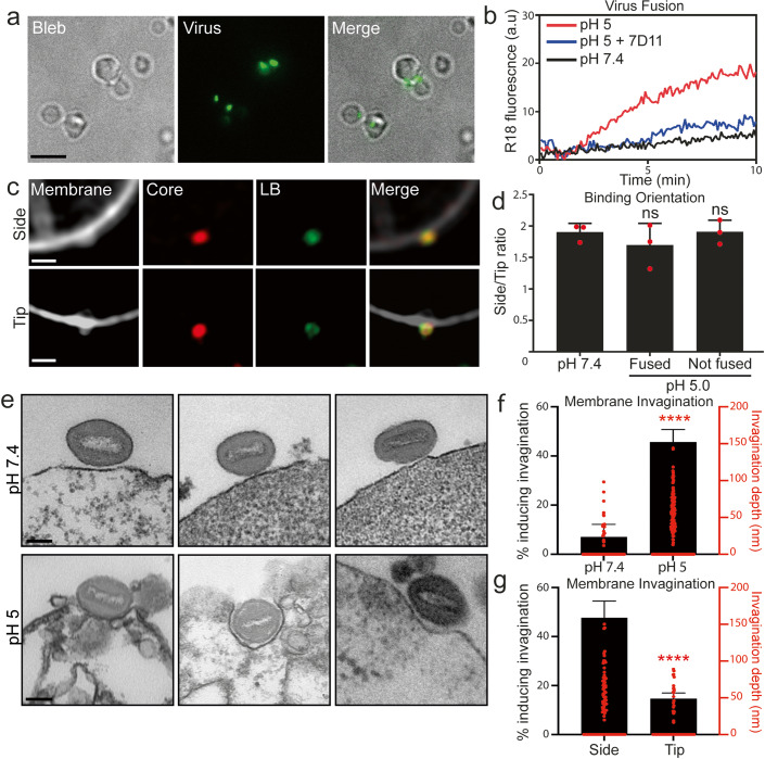
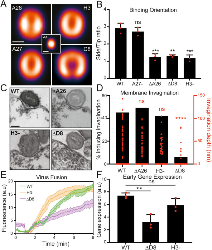
Cellular attachment of viruses determines their cell tropism and species specificity. For entry, vaccinia, the prototypic poxvirus, relies on four binding proteins and an eleven-protein entry fusion complex. The contribution of the individual virus binding proteins to virion binding orientation and membrane fusion is unclear. Here, we show that virus binding proteins guide side-on virion binding and promote curvature of the host membrane towards the virus fusion machinery to facilitate fusion. Using a membrane-bleb model system together with super-resolution and electron microscopy we find that side-bound vaccinia virions induce membrane invagination in the presence of low pH. Repression or deletion of individual binding proteins reveals that three of four contribute to binding orientation, amongst which the chondroitin sulfate binding protein, D8, is required for host membrane bending. Consistent with low-pH dependent macropinocytic entry of vaccinia, loss of D8 prevents virion-associated macropinosome membrane bending, disrupts fusion pore formation and infection. Our results show that viral binding proteins are active participants in successful virus membrane fusion and illustrate the importance of virus protein architecture for successful infection.

Keywords: Glycosaminoglycans; Membrane Bending; Membrane Fusion; Poxvirus; Virus Entry.


Conflict of interest statement

The authors declare no competing interests.

Figures

Figure 1
Figure 1. VACV binds to, fuses with and mediates curvature of membrane blebs.
(A) Blebs incubated with A4-EGFP virus at 4 °C for 1 h, washed and imaged. Scale bar, 5 μm. (B) VACV hemifusion rates (R18 dequenching assay) with blebs at pH 7.4 or pH 5.0. 7D11 fusion neutralizing antibody was used as a positive control. Fluorescence was normalized to the initial value and fully de-quenched value upon TX-100 addition. (C) Examples SIM images of mCherry-A4 EGFP-F17 VACV bound to blebs in a side-on (upper row) and tip-on (lower row) orientation. Scale bar = 1 µm. (D) Quantification of side/tip binding ratio at different pH’s and fusion states (n = 50 virions/replicate). (E) TEM images of VACV bound to HeLa cell derived blebs at pH 7.4 (top row) or pH 5.0 (bottom row). Scale bar = 100 nm. (F) Quantification of membrane invagination depth under virions at pH 7.4 and pH 5.0 (n > 50 virions/replicate). (G) Quantification of side-on vs. tip-on bound virion invagination (n > 50 virions/replicate). Data information: In (D, F, G), data are mean ± standard deviation (SD) of biological triplicates. Statistical analysis was performed using unpaired two-tailed t-tests (****P < 0.0001; ns, not significant (P > 0.05)). Source data are available online for this figure.
Figure 2
Figure 2. VACV binding protein D8 is required for host membrane invagination and fusion.
(A) VirusMapper localization models of VACV binding proteins. EGFP-A4 core (center) was used to correlate virion orientation. Models are representative of n > 180 virions. Scale bar, 200 nm. (B) Analysis of side/tip binding ratio of WT and binding protein mutants on blebs (n = 50 virions/replicate). (C) TEM images of WT and binding protein mutants bound to HeLa blebs at pH 5.0. Scale bar, 100 nm. (D). Quantification of percent invagination and invagination depth of binding protein mutants at pH 5.0 (n > 50 virions/mutant). (E) Hemifusion rates of WT, ΔD8 and H3- virions on HeLa cells using the R18 dequenching assay. (F) Comparison of WT, ΔD8 and H3- virus early gene expression at 2 hpi by RT-qPCR of early gene C11R. Data information: For (B, E), data are means ± SEM of biological triplicates and for (F) data are means ± SD. Statistical analysis was performed using unpaired two-tailed t tests (****P < 0.0001; ***P < 0.001; **P < 0.01; ns, not significant (P > 0.05)). Source data are available online for this figure.
Figure 3
Figure 3. D8-mediated invagination is a critical component of VACV low pH-dependent fusion.
(A) Representative TEM images of WT and A28- virions bound to HeLa blebs at pH 5.0. Scale bar, 100 nm. (B) Quantification of WT and A28- virion % invagination and invagination depth comparing under pH 5.0 conditions (from (A)) (n = 80 virions/mutant). (C) Representative TEM images of A28- or A28-ΔD8 virions within macropinosomes. Scale bar, 100 nm. (D) Quantification of curvature of the macropinosome membrane around individual A28- or A28-ΔD8 virions (from (F)) using the Fiji plugin Kappa (n > 60 virions/mutant). (E) Model of low-pH mediated D8-mediated macropinosome membrane curvature and VACV fusion. Data information: Statistical analysis was performed using unpaired two-tailed t tests (****P < 0.0001; ns, not significant (P > 0.05)). Source data are available online for this figure.
Figure 4
Figure 4. D8-mediated invagination requires pH sensitive chondroitin sulfate binding.
(A) TEM images of WT VACV bound to HS + CS+ or HS-CS- cell-derived blebs at pH 5.0. Scale bar = 100 nm. (B) Quantification of % invagination and invagination depth of virions from A (n > 60 virions/mutant). (C) Hemifusion rates of WT virus on HS + CS+ and HS-CS- cells using the R18 dequenching assay. (D) TEM images of WT VACV bound to HS + CS+ or HS-CS+ cell-derived blebs at pH 5.0. Scale bars = 100 nm. (E) Quantification of % invagination and invagination depth of virions in (D) (n = 95 virions/mutant). (F) Hemifusion rates of WT virus on HS + CS+ and HS-CS+ cells using the R18 dequenching assay. (G) Immunoprecipitation analysis of D8 binding to biotinylated CS-E or CS-A. (H) Immunoprecipitation analysis of D8 binding to biotinylated CS-E at neutral (7.4) and low (5.0) pH. (I) Digestion of D8 from the surface of WT virions at neutral (7.4) and low (5.0) pH using varying concentrations of papain. Data information: (C, F) data are means ± SEM. GI were performed in triplicate and representative blots shown. Statistical analysis was performed using unpaired two-tailed t tests (****P < 0.0001; ns, not significant (P > 0.05)). Source data are available online for this figure.
Figure EV1
Figure EV1. Cell-derived membrane blebs as a minimal cell system.
(A) To prepare blebs Latrunculin B is added to cells to induce blebbing. Cells are shaken to detach blebs, cellular debris removed by a slow spin step (300 × g), blebs collected by a fast spin step (4000 × g), and remaining large debris removed by filtration through a 5 μm pore filter. (B) Representative brightfield (LHS, scale bar; 1 µm) and TEM (RHS, scale bar; 500 nm) images of blebs after purification. (C) Histogram of bleb diameter range. (D) Blebs were scored for mono-, bi- and multi-lobulation (n > 100 blebs/repeat). (E) Blebs were stained for actin (green) and the plasma membrane (PM; magenta). Scale bar, 5 µm. (F) Bleb cortical actin was reconstituted (R) with ATP or not (NR), stained for both actin and the PM and the percentage of blebs with an actin cortex calculated (n > 100 blebs/repeat). (G) Stability of the actin cortex over time at 37 °C between NR and R blebs was determined by intensity measurements of the actin stain on z-projections (n > 40 blebs). Data information: For (D, F, G), data are means ± SD. Statistical analysis was performed using unpaired two-tailed t tests (ns, not significant (P > 0.05)).
Figure EV2
Figure EV2. VACV binding proteins reside at virion sides and are differentially required for VACV binding.
(A) Quantification of VACV binding protein polarity factors using data from the models in Fig. 2A. A polarity factor of less than one corresponds to concentration of the protein at the sides of MVs (n = 50 virions per repeat). (B) Representative STORM images of VACV binding proteins on individual MVs. Scale bar = 200 nm. (C) Binding affinities of WT and recombinant binding protein mutant VACVs on HS + CS+ cells (n = 4 biological repeats). (D) Representative immunoblot of D8 protein packaging in A28- and A28-ΔD8 virions. Molecular weight markers are indicated at right. Data information: (A, C) data are means ± SD. Statistical analysis was performed using unpaired two-tailed t tests (***P < 0.001; **P < 0.01; ns, not significant (P > 0.05)).
Figure EV3
Figure EV3. CS-E is the major GAG used by VACV for binding.
(A) Binding affinities of WT VACV on HS + CS + , HS-CS+ and HS-CS- cells. (B) SIM images of VACV bound to HS + CS + , HS-CS+ and HS-CS- derived blebs scale bar = 2 µm. A4-EGFP VACV (magenta) and PM (green). (C) Quantification of % invagination and invagination depth of WT and ∆D8 virions on HS + CS+ cells (n > 60 virions/mutant). (D) Binding affinities of WT VACV with GAG pre-incubation on HS + CS+ cells. (E) Binding affinities of WT virus with CS-E preincubation on HS + CS+ and HS-CS+ cells. Data is normalized to no CS-E preincubation on the given cell type. (F) Binding affinities of A27- virions with GAG pre-incubation on HS + CS+ cells. Data information: (A, DF) data are means ± SD. Statistical analysis was performed using unpaired two-tailed t tests (***P < 0.001; **P < 0.01; *P < 0.05; ns, not significant (P > 0.05)).

References

    1. Aldaz-carroll L, Whitbeck JC, De Leon MP, Lou H, Hirao L, Isaacs SN, Moss B, Eisenberg RJ, Cohen GH. Epitope-mapping studies define two major neutralization sites on the vaccinia virus extracellular enveloped virus. J Virol. 2005;79:6260–6271. doi: 10.1128/JVI.79.10.6260-6271.2005. - DOI - PMC - PubMed
    1. Banfield BW, Leduc Y, Esford L, Schubert K, Tufaro F. Sequential isolation of proteoglycan synthesis mutants by using herpes simplex virus as a selective agent: evidence for a proteoglycan-independent virus entry pathway. J Virol. 1995;69:3290–3298. doi: 10.1128/jvi.69.6.3290-3298.1995. - DOI - PMC - PubMed
    1. Bengali Z, Satheshkumar PS, Moss B. Orthopoxvirus species and strain differences in cell entry. Virology. 2013;433:506–512. doi: 10.1016/j.virol.2012.08.044. - DOI - PMC - PubMed
    1. Bengali Z, Townsley AC, Moss B. Vaccinia virus strain difference in cell attachment and entry. Virology. 2010;389:132–140. doi: 10.1016/j.virol.2009.04.012. - DOI - PMC - PubMed
    1. Bergefall K, Trybala E, Johansson M, Uyama T, Naito S, Yamada S, Kitagawa H, Sugahara K, Bergstro T. Chondroitin sulfate characterized by the E-disaccharide unit is a potent inhibitor of herpes simplex virus infectivity and provides the virus binding sites on gro2C cells. J Biol Chem. 2005;280:32193–32199. doi: 10.1074/jbc.M503645200. - DOI - PubMed