Clinical Trial
. 2024 Mar;30(3):762-771.
doi: 10.1038/s41591-024-02805-1. Epub 2024 Feb 6.

MYC targeting by OMO-103 in solid tumors: a phase 1 trial

Affiliations
Clinical Trial

MYC targeting by OMO-103 in solid tumors: a phase 1 trial

Elena Garralda et al. Nat Med. 2024 Mar.

Abstract

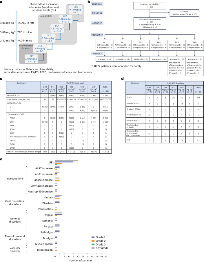
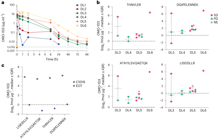
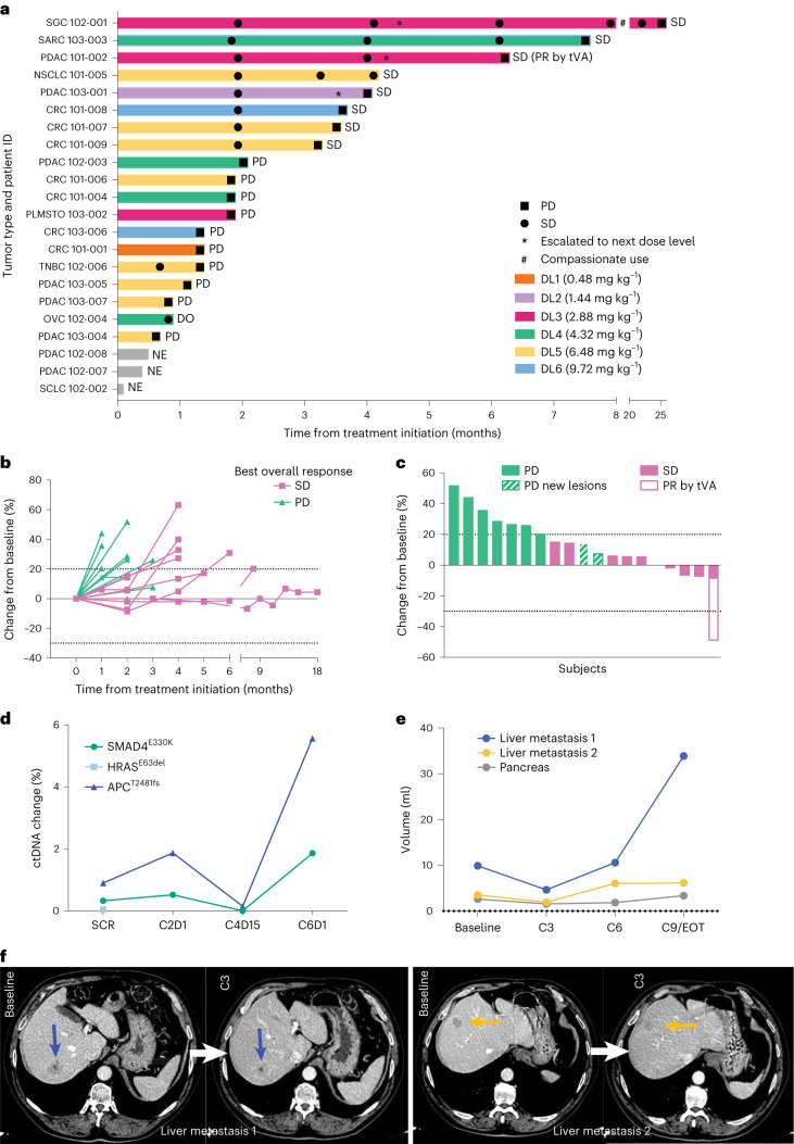
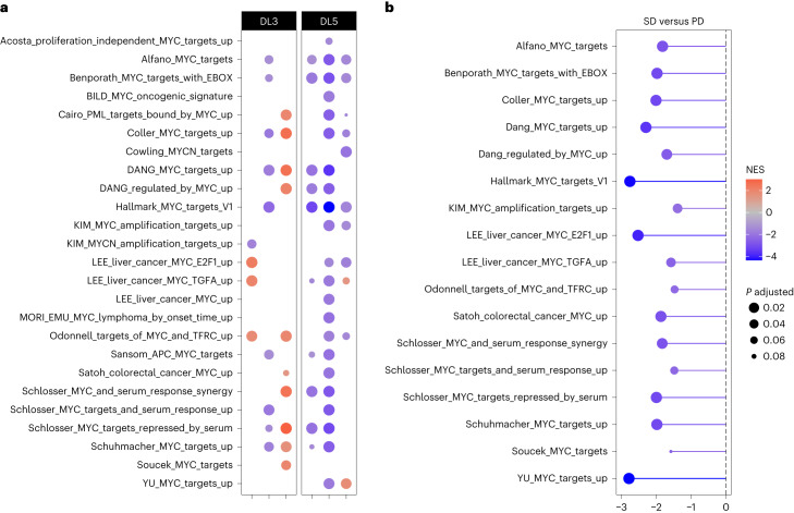
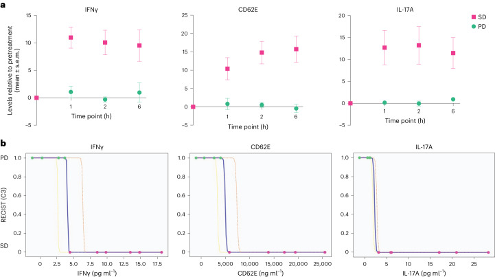
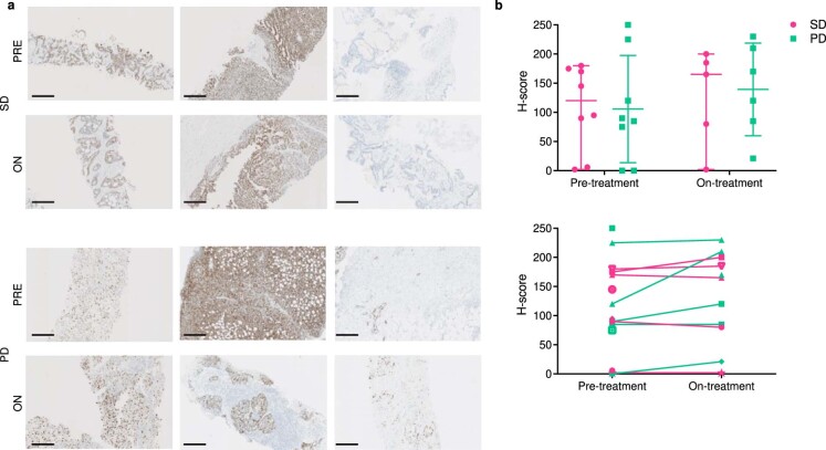
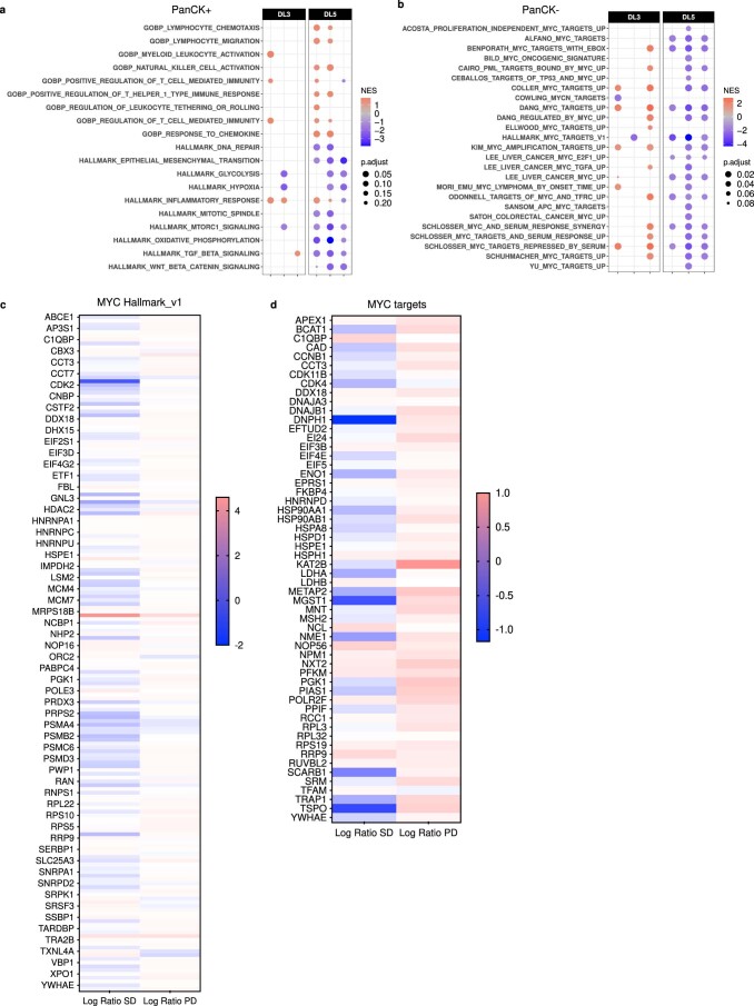
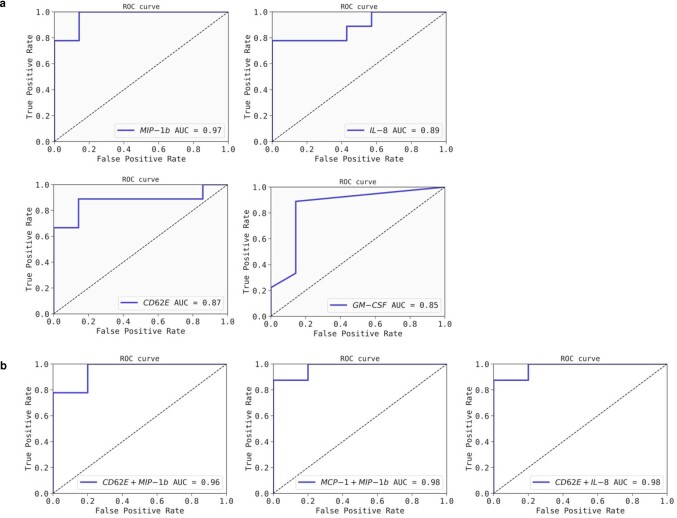
Among the 'most wanted' targets in cancer therapy is the oncogene MYC, which coordinates key transcriptional programs in tumor development and maintenance. It has, however, long been considered undruggable. OMO-103 is a MYC inhibitor consisting of a 91-amino acid miniprotein. Here we present results from a phase 1 study of OMO-103 in advanced solid tumors, established to examine safety and tolerability as primary outcomes and pharmacokinetics, recommended phase 2 dose and preliminary signs of activity as secondary ones. A classical 3 + 3 design was used for dose escalation of weekly intravenous, single-agent OMO-103 administration in 21-day cycles, encompassing six dose levels (DLs). A total of 22 patients were enrolled, with treatment maintained until disease progression. The most common adverse events were grade 1 infusion-related reactions, occurring in ten patients. One dose-limiting toxicity occurred at DL5. Pharmacokinetics showed nonlinearity, with tissue saturation signs at DL5 and a terminal half-life in serum of 40 h. Of the 19 patients evaluable for response, 12 reached the predefined 9-week time point for assessment of drug antitumor activity, eight of those showing stable disease by computed tomography. One patient defined as stable disease by response evaluation criteria in solid tumors showed a 49% reduction in total tumor volume at best response. Transcriptomic analysis supported target engagement in tumor biopsies. In addition, we identified soluble factors that are potential pharmacodynamic and predictive response markers. Based on all these data, the recommended phase 2 dose was determined as DL5 (6.48 mg kg-1).ClinicalTrials.gov identifier: NCT04808362 .


Conflict of interest statement

E.G. is Consultant-Advisor for Roche/Genentech, F.Hoffmann/La Roche, Ellipses Pharma, Neomed Therapeutics1, Inc., Boehringer Ingelheim, Janssen Global Services, SeaGen, TFS, Alkermes, Thermo Fisher, Bristol-Myers Squibb, MabDiscovery, Anaveon, F-Star Therapeutics and Hengrui. L.S. is Consultant and CEO of Peptomyc. V.M. is Consultant of Roche, Bayer, BMS, Janssen and Basilea. E.C. is Consultant-Advisor for Nanobiotix, Janssen-Cilag, Roche-Genentech, TargImmune Therapeutics, Servier, Bristol-Myers Squibb, Amunix, Adcendo, Anaveon, AstraZeneca/Medimmune, Chugai Pharma, MonTa, MSD Oncology, Nouscom, OncoDNA, T-Knife, Elevation Oncology, PharmaMar, Ellipses Pharma, Syneos Health, Genmab and Diaccurate. Employment. M.-E.B., S.C.-S., S.M.-M., L.F., S.L.-E., V.C.C., J.M., M.N. and L.S. are employees of Peptomyc. E.C. is an employee of START. Personal financial interests: L.S. and M.-E.B. are cofounders and shareholders of Peptomyc and inventors of patent application no. WO2014180889 A8 that covers the use of the Omomyc miniprotein in medicine, held by VHIO and licenced to Peptomyc. S.C.-S., S.L.-E., L.F., J.R.W. and M.N. are also shareholders of Peptomyc. E.G. performs research for Novartis, Roche, Thermo Fisher, AstraZeneca, Taiho and BeiGene; she is in Speakers Bureau for Merck Sharp & Dohme, Roche, Thermo Fisher, Lilly and Novartis; she runs clinical trials as PI or Co-PI with Agios Pharmaceuticals, Amgen, Bayer, Beigene USA, Blueprint Medicines, BMS, Cellestia Biotech, Debiopharm, F.Hoffmann/La Roche, Ltd, Forma Therapeutics, Genentech, Inc., Genmab B.V., GSK, Glycotope Gmbh, Incyte Biosciences, Incyte Corporation, ICO, Kura Oncology, Inc, Lilly, S.A, Loxo Oncology, Inc, Macrogenics, Inc, Menarini Ricerche Spa, Merck, Sharp & Dohme de España, S.A, Nanobiotix, S.A, Novartis Farmacéutica, S.A, Pfizer, SLU, Pharma Mar, S.A.U, Pierre Fabre Medicament, Principia Biopharma, Inc., Psioxus Therapeutics, Ltd, Sanofi, Sierra Oncology, Inc, Sotio A.S and Symphogen A/S. V.M. is Principal Investigator and receives institutional funding from AbbVie, AceaBio, Adaptimmune, ADC Therapeutics, Aduro, Agenus, Amcure, Amgen, Astellas, AstraZeneca Bayer Beigene BioInvent International AB, BMS, Boehringer, Boheringer, Boston, Celgene, Daichii Sankyo, DEBIOPHARM, Eisai, e-Terapeutics, Exelisis, Forma Therapeutics, Genmab, GSK, Harpoon, Hutchison, Immutep, Incyte, Inovio, Iovance, Janssen, Kyowa Kirin, Lilly, Loxo, MedSir, Menarini, Merck, Merus, Millennium, MSD, Nanobiotix, Nektar, Novartis, Odonate Therapeutics, Pfizer, Pharma Mar, PharmaMar, Principia, PsiOxus, Puma, Regeneron, Rigontec, Roche, Sanofi, Sierra Oncology, Synthon, Taiho, Takeda, Tesaro, Transgene, Turning Point Therapeutics and Upshersmith. E.C. is Principal Investigator and receives institutional funding from START, Pharma Mar, EORTC, Sanofi, BeiGene, Novartis and Merus N.V.; he is a shareholder of START and Oncoart Associated and receives honoraria from HM Hospitales Group; he receives research funding from START; he is President and Founder of Foundation Investigational Therapeutics in Oncological Sciences; and he has a not-for-profit relationship with PharmaMar and the CRIS Cancer Foundation. The other authors declare no competing interests.

Figures

Fig. 1
Fig. 1. Overall trial design and safety.
a, Schematic of dose-escalation design. PAD, pharmacologically active dose; TED, therapeutically effective dose; NOAEL, no observed adverse effect level; n, number of patients. b, CONSORT flow diagram. c, Demographic and baseline characteristics. ECOG PS, ECOG performance status. d, Overall safety. TEAE, treatment-emergent adverse event. e, TRAEs (n = 22). System organ class and symptoms are shown. SCLC, small cell lung cancer; NSCLC, non-small cell lung cancer; TNBC, triple-negative breast cancer; ALAT, alanine aminotransferase; ASAT, aspartate aminotransferase. Source data
Fig. 2
Fig. 2. PK of OMO-103 in serum and detection in patient biopsies.
a, PK profiles at different DLs following a single intravenous infusion of OMO-103 (n = 22). LLOQ, lower limit of quantification. Statistical average value and standard deviation are shown. b, Quantification of functional Omomyc by MS in FFPE on-treatment biopsies from MYCure patients for DL3–6 following administration of at least three intravenous infusions of OMO-103 (n = 12). The sequence of peptides used for detection of the Omomyc protein is indicated on top of the x axis. Median value and interquartile range (IQR) are shown. c, Results from end-of-treatment (EOT) biopsy of a patient with PDAC from DL3 who remained for >6 months in the study and had received their last infusion 19 days before biopsy, compared with those obtained at cycle 1, day 16 (C1D16). NE, not evaluable. Source data
Fig. 3
Fig. 3. Evaluation of tumor response in all cohorts and time-dependent disease evaluation in one patient with PDAC.
a, Swimmer plot of 22 enrolled patients. SD, stable disease; PD, progressive disease; DO, dropout; PR, partial response; tVA, Total volumetric analysis. b, Spider plot showing changes in target lesion volume from 19 evaluable patients. c, Waterfall graph indicating change in volume of target lesions (RECIST v.1.1) at best response of 19 evaluable patients. d, ctDNA dynamics of the three somatic alterations identified by Guardant360. e,f, Baseline, C3, C6 and C9/EOT volume measurement of individual lesions by CT scan (e) and representative images (f). Blue and yellow arrows indicate lesions quantified in e.
Fig. 4
Fig. 4. Target engagement analysis by DSP.
a,b, In DL3 and 5, three paired biopsies each were analyzed (n = 6 in total). a, GSEA comparing the status of each MYC gene set in on-treatment (C1D15)** versus pretreatment biopsies. Normalized enrichment score (NES) is represented by a color scale from red (enriched post treatment) to blue (enriched in pretreatment compared with post), while adjusted P value is represented by circle size. For each patient, three to five regions of interest were compiled. b, Lollipop graph representing comparison by GSEA of the status of MYC gene sets in samples from patients with SD versus PD. NES is represented by the length of the lollipop and color scale, while the adjusted P value is represented by circle size.
Fig. 5
Fig. 5. Patients that clinically benefited from OMO-103 showed low baseline levels of MIP-1β, IL-8, CD62E and GM-CSF, with model pairings of different soluble factors providing outstanding outcome predictors.
ac, Levels of soluble factors were measured in patient serum samples at pretreatment using the Luminex technique. a,b, Nine patients with PD and seven with SD were included in the analyses. a, Patients showing disease stabilization at cycle 3 displayed significantly lower levels of MIP-1β, IL-8, CD62E and GM-CSF compared with those with PD (median shown). A two-sided Mann–Whitney U-test, with no adjustment for multiple comparisons, was used for statistical analysis. b, Binomial logistic regression analysis of the association of cytokine and chemokine levels at pretreatment with the probability of disease stabilization. ORs of soluble factors in patients are shown in the forest plot (left) and summary table (right); mean and CI are shown. c, Soluble factor combination models generated using QLattice technology; data from nine patients with PD and six with SD were used to generate the models. Plots of combination models CD62E + MIP-1β, MCP-1 + MIP-1β and CD62E + IL-8 are shown in relationship to patient response. SD and PD are indicated by pink and green dots, respectively. Colored lines correspond to confidence bands: blue indicates the mean, orange 95% CI and yellow 5% CI. Patient no. 101-009 is marked with a rhombus because their data were not available at the time of generation of the models and were later included. Source data
Fig. 6
Fig. 6. Following OMO-103 infusion, patients with disease stabilization at C3 showed significantly increased levels of IFNγ, CD62E and IL-17A, which can be used to identify patients with SD or PD.
a,b, Levels of different soluble factors were measured in patient serum samples taken at different time points following OMO-103 treatment and were determined using the Luminex technique. Four and six patients with PD and SD, respectively, were included in the analysis. a, At the onset of C3, patients with disease stabilization at the following CT scan showed a significant increase in levels of IFNγ (P = 0.000148), CD62E (P = 0.000338) and IL-17A (P = 0.0013) following OMO-103 infusion compared with patients with PD. Mean and s.e.m. are shown. A two-sided Welch’s t-test was used with Bonferroni correction to adjust for multiple comparisons. b, Individual IFNγ, CD62E and IL-17A models generated with QLattice technology. Maximum serum levels of the indicated cytokines were used to generate the models. Serum levels of patients with PD and SD are indicated as 1 and 0, respectively. Colored lines correspond to confidence bands: blue indicates the mean, yellow 5% CI and orange 95% CI. Source data
Extended Data Fig. 1
Extended Data Fig. 1. MYC levels in patient biopsies.
(a) Representative images of pre- and on-treatment biopsies from stable and progressive disease patients. (b) Quantification of MYC protein levels determined by immunohistochemistry and given as H-score, a semi-quantitative assessment that takes into account the intensity (graded as 0, 1+, 2+, or 3+ for each cell) and percentage of positive cells according to the formula: H-score = [1 × (% cells 1+) + 2 × (% cells 2+) + 3 × (% cells 3+)]. n = 16 patients at pre-treatment. n = 11 patients at on-treatment. In the upper plot, the median value and 95% confidence intervals are shown. In the lower plot, the variation in H-score between pre- and on-treatment is shown per patient, when possible. A mixed-effects analysis with Bonferroni’s correction for multiple tests was used.
Extended Data Fig. 2
Extended Data Fig. 2. DSP assessment of modulation of additional pathways in the tumor related to cancer and the immune system and target engagement in PanCK- cells, and proteomic analysis of MYC targets.
3 paired biopsies were analyzed in DL3 and 3 in DL5 (n=6 in total). GSEA comparing in on-treatment versus pre-treatment biopsies the (a) status of immune- and cancer-related gene sets in the PanCK+ segment and (b) status of each MYC gene set in the PanCK- segment. The NES is represented by a color scale from red (enriched post-treatment) to blue (enriched in the pre-treatment compared to the post), while the adjusted p-value is represented by the size of each circle. For each patient, 3–5 regions of interest were compiled. (c and d) 2 paired biopsies were analyzed in DL3 and 2 in DL5 (n=4 in total) by UltraDeep Protein Profiling. The Log Ratio of SD and PD patients was calculated per protein by dividing on-treatment by pre-treatment values from a subset of (c) 179 proteins from the MYC Hallmarks v1 and (d) 56 direct targets of MYC. Such values are represented by a color scale from red (upregulated) to blue (downregulated).
Extended Data Fig. 3
Extended Data Fig. 3. Receiver Operating Characteristic (ROC) curve analysis of the identified soluble factors and predictive combination models.
(a) Receiver Operating Characteristic (ROC) curve analysis for individual soluble factors to predict response to OMO-103. Individual ROC curve graphs and their corresponding Area Under the Curve (AUC) scores for each individual soluble factor are shown. Each ROC curve illustrates the relationship between the true positive rate and the false positive rate across various classification thresholds. (b) ROC curve analysis and ROC-AUC scores of the predictive combination models are shown below. ROC-AUC scores of the combination models are higher than those of the individual factors, suggesting an increased predictive power.
Extended Data Fig. 4
Extended Data Fig. 4. Receiver Operating Characteristic (ROC) curve analysis of the individual pharmacodynamic models.
Receiver Operating Characteristic (ROC) curve analysis of the individual pharmacodynamic models to identify SD vs PD patients upon OMO-103 treatment. Individual ROC curve graphs and ROC-AUC scores for each individual model are shown. Each ROC curve illustrates the relationship between the true positive rate and the false positive rate across various classification thresholds.
Extended Data Fig. 5
Extended Data Fig. 5. The pharmacodynamic signature can be associated to maintained SD and is lost upon disease progression.
The levels of IFN-γ, CD62E and IL-17A are not transiently increased after OMO-103 infusion for patients 101-002 and 103-003 at C9, time point at which they displayed PD as per RECIST criteria. Tables indicate the values relative to pre-dose at the maximum peak, how the model classifies them in terms of SD or PD, and the probability to become PD according to their maximum relative levels. Graphs of the three independent models show the relative values of each soluble factor at C3 and C9. C3 is marked as a red dot and C9 as a yellow dot. CI: confidence interval.

Comment in

  • MYC inhibitor achieves phase I success.
    Crunkhorn S. Crunkhorn S. Nat Rev Drug Discov. 2024 Apr;23(4):253. doi: 10.1038/d41573-024-00042-2. Nat Rev Drug Discov. 2024. PMID: 38448668 No abstract available.

References

    1. Dang CV, Reddy EP, Shokat KM, Soucek L. Drugging the ‘undruggable’ cancer targets. Nat. Rev. Cancer. 2017;17:502–508. doi: 10.1038/nrc.2017.36. - DOI - PMC - PubMed
    1. Dhanasekaran R, et al. The MYC oncogene – the grand orchestrator of cancer growth and immune evasion. Nat. Rev. Clin. Oncol. 2022;19:23–36. doi: 10.1038/s41571-021-00549-2. - DOI - PMC - PubMed
    1. Stine ZE, Walton ZE, Altman BJ, Hsieh AL, Dang CV. MYC, metabolism, and cancer. Cancer Discov. 2015;5:1024–1039. doi: 10.1158/2159-8290.CD-15-0507. - DOI - PMC - PubMed
    1. Nie Z, et al. Dissecting transcriptional amplification by MYC. eLife. 2020;9:e52483. doi: 10.7554/eLife.52483. - DOI - PMC - PubMed
    1. Whitfield JR, Beaulieu ME, Soucek L. Strategies to inhibit Myc and their clinical applicability. Front. Cell Dev. Biol. 2017;5:10. doi: 10.3389/fcell.2017.00010. - DOI - PMC - PubMed

Publication types

Associated data