. 2024 Feb;13(2):e12409.
doi: 10.1002/jev2.12409.

Identification of specific markers for human pluripotent stem cell-derived small extracellular vesicles

Affiliations

Identification of specific markers for human pluripotent stem cell-derived small extracellular vesicles

Zhengsheng Chen et al. J Extracell Vesicles. 2024 Feb.

Erratum in

Abstract

Pluripotent stem cell-derived small extracellular vesicles (PSC-sEVs) have demonstrated great clinical translational potential in multiple aging-related degenerative diseases. Characterizing the PSC-sEVs is crucial for their clinical applications. However, the specific marker pattern of PSC-sEVs remains unknown. Here, the sEVs derived from two typical types of PSCs including induced pluripotent stem cells (iPSC-sEVs) and embryonic stem cells (ESC-sEVs) were analysed using proteomic analysis by liquid chromatography with tandem mass spectrometry (LC-MS/MS), and surface marker phenotyping analysis by nanoparticle flow cytometry (NanoFCM). A group of pluripotency-related proteins were found to be enriched in PSC-sEVs by LC-MS/MS and then validated by Western Blot analysis. To investigate whether these proteins were specifically expressed in PSC-sEVs, sEVs derived from seven types of non-PSCs (non-PSC-sEVs) were adopted for analysis. The results showed that PODXL, OCT4, Dnmt3a, and LIN28A were specifically enriched in PSC-sEVs but not in non-PSC-sEVs. Then, commonly used surface antigens for PSC identification (SSEA4, Tra-1-60 and Tra-1-81) and PODXL were gauged at single-particle resolution by NanoFCM for surface marker identification. The results showed that the positive rates of PODXL (>50%) and SSEA4 (>70%) in PSC-sEVs were much higher than those in non-PSC-sEVs (<10%). These results were further verified with samples purified by density gradient ultracentrifugation. Taken together, this study for the first time identified a cohort of specific markers for PSC-sEVs, among which PODXL, OCT4, Dnmt3a and LIN28A can be detected with Western Blot analysis, and PODXL and SSEA4 can be detected with NanoFCM analysis. The application of these specific markers for PSC-sEVs identification may advance the clinical translation of PSCs-sEVs.

Keywords: pluripotent stem cells; single-particle resolution; small extracellular vesicles; specific markers.


Conflict of interest statement

Yang Wang holds research grants from Shanghai EOOXOM Biotechnology Co., Ltd. Other authors declare no conflict of interest.

Figures
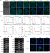
FIGURE 1
FIGURE 1
Characterization of ESC and iPSC. One ESC line (H9) and one iPSC line (nciPS01) were selected for sEVs collection. (a) Representative bright‐field images and fluorescent images of PSC markers including OCT4, SOX2, NANOG, SSEA4, TRA‐1‐60, and TRA‐1‐81 of ESC and iPSC. Nucleus was stained with DAPI. Scale bar, 100 μm. (b) Flow cytometry analysis of PSC markers including OCT4, NANOG, SSEA4, TRA‐1‐60, and TRA‐1‐81. (c) RT‐PCR for pluripotency transcripts of ESC and iPSC. (d) RT‐PCR analysis of various differentiation markers for the three germ layers of PSC formed‐EBs. (e) Immunofluorescence detection of cells positive for TUJ1 (ectoderm), T (mesoderm), and FOXA2 (endoderm). Scale bar, 50 μm. ESC, embryonic stem cells; iPSC, induced pluripotent stem cells; EB, Embryoid body.
FIGURE 2
FIGURE 2
Characterization of PSC‐sEVs. The morphology, particle size, classical protein markers, yields in terms of particles and proteins, and surface potential of PSC‐sEVs were detected. (a) Representative TEM images of ESC‐sEVs and iPSC‐sEVs. Scale bar, 200nm. (b) Representative profiles of size distribution of ESC‐sEVs and iPSC‐sEVs determined by NanoFCM. (c) Representative bands of Western Blot for sEVs markers (CD9, CD63, Alix, and TSG101) and non‐sEVs markers (Calnexin and GM130) in PSCs and PSC‐sEVs. (d‐f) Evaluation of ESC‐sEVs and iPSC‐sEVs yield in terms of the particle concentration and the protein concentration (n=5). (g) Zeta potential of ESC‐sEVs and iPSC‐sEVs (n=10). PSC‐sEVs, pluripotent stem cell‐derived sEVs; TEM, transmission electron microscope; ESC‐sEVs, embryonic stem cell‐derived sEVs; iPSC‐sEVs, induced pluripotent stem cell‐derived sEVs.
FIGURE 3
FIGURE 3
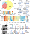
Proteomics analysis of PSC‐sEVs and Western Blot validation of pluripotency‐related proteins. LC‐MS/MS analysis and subsequent bioinformatics analysis were applied to identify the protein contents of PSC‐sEVs. Candidate proteins were selected for Western Blot validation. (a) Venn diagram showing the overlapping and unique proteins in ESC‐sEVs and iPSC‐sEVs with Vesiclepedia database. (b‐d) Gene Ontology cellular component, molecular function, and biological process enrichment analysis of proteins identified in ESC‐sEVs and iPSC‐sEVs. Upper y axis, ‐log (p‐value); lower y axis, percentage. (e‐f) Heatmap of corrected Log2 LFQ intensity showing the common sEVs‐related markers (e) and pluripotency‐related markers (f) in proteomes of ESC‐sEVs and iPSC‐sEVs. White rectangle, not exist in the proteome. (g‐h) Western Blot analysis of the putative markers, including PODXL, YB1, IFITM1, LIN28A, OCT4, Dnmt3A, and CDH1, in PSC and PSC‐sEVs. *, p <0.05; **, p <0.01; #, p <0.0001. LC‐MS/MS, liquid chromatography with tandem mass spectrometry; PSC‐sEVs, pluripotent stem cell‐derived sEVs; ESC‐sEVs, embryonic stem cell‐derived sEVs; iPSC‐sEVs, induced pluripotent stem cell‐derived sEVs; LFQ, label‐free quantification.
FIGURE 4
FIGURE 4
Validation of specificity of the markers with Western Blot. Seven non‐PSC cell lines derived sEVs were used for validation of the specificity of the putative markers for PSC‐sEVs. (a‐b) Representative bands of Western Blot showing that PODXL, LIN28A, OCT4, and Dnmt3A were specifically detected in PSC‐sEVs, while YB1 and IFITM1 were also detected in normal cell lines derived sEVs. Statistical analysis was performed for semi‐quantification of Western Blot results. #, p <0.0001. (c‐d) Representative bands of Western Blot showing that LIN28A, OCT4, and Dnmt3A were specifically detected in PSC‐sEVs, while PODXL, YB1, and IFITM1 were also detected in tumor cell lines derived sEVs. Statistical analysis was performed for semi‐quantification of Western Blot results. #, p <0.0001. PSC‐sEVs, pluripotent stem cell‐derived sEVs.
FIGURE 5
FIGURE 5
Evaluation of surface markers of PSC‐sEVs at single particle‐resolution. Three sEVs traditional surface markers (CD9, CD63, and CD81), one validated protein (PODXL), and three specific surface antigens on PSC (SSEA4, Tra‐1‐60, and Tra‐1‐81) were selected for surface marker detection. (a‐d) NanoFCM analysis for the sEVs traditional surface markers including CD9, CD63, CD81, and combination of CD9/CD63/CD81 on PSC‐sEVs. Left panel, histograms showing the fluorescence intensity. Right panel, bar graphs showing the positive rates of surface markers. (e‐h) NanoFCM analysis for the indicated surface markers including PODXL, SSEA4, Tra‐1‐60, and Tra‐1‐81 on PSC‐sEVs. Left panel, histograms showing the fluorescence intensity. Right panel, bar graphs showing the positive rates of surface markers. PSC‐sEVs, pluripotent stem cell‐derived sEVs.
FIGURE 6
FIGURE 6
Validation of specificity of the markers with Immunofluorescent staining at single particle‐resolution. Seven non‐PSC cell lines derived sEVs were used for validation of specificity of the putative surface markers for PSC‐sEVs. (a‐d) Marker phenotyping analysis with NanoFCM for the sEVs traditional surface markers including CD9, CD63, CD81 and combination of CD9/CD63/CD81 on sEVs. Left panel, histograms showing the fluorescence intensity. Right panel, bar graphs showing the positive rates of surface markers. (e‐f) Marker phenotyping analysis with NanoFCM for the PODXL and SSEA4 on sEVs, showing the positive rates of PODXL and SSEA4 in non‐PSC‐sEVs were much lower than those in PSC‐sEVs. Left panel, histograms showing the fluorescence intensity. Right panel, bar graphs showing the positive rates of surface markers. PSC‐sEVs, pluripotent stem cell‐derived sEVs.
FIGURE 7
FIGURE 7
Validation of sEVs preparations via DGUC. UC samples of PSC‐sEVs were further purified with DGUC and then analyzed with Western Blot and NanoFCM. (a) Workflow of sucrose density gradient ultracentrifugation. (b) Western Blot analysis of TSG101 in PSC‐sEVs from the distinct DGUC fractions showing that sEVs were enriched in F6‐F8. (c‐d) Western Blot analysis of the putative markers, including PODXL, LIN28A, OCT4, and Dnmt3A, in PSC and PSC‐sEVs preparations via DGUC. **, p <0.01; ***, p <0.001; #, p <0.0001. ns, no significant difference. (e‐j) Marker phenotyping analysis with NanoFCM for the sEVs surface markers including CD9, CD63, CD81, combination of CD9/CD63/CD81, PODXL, and SSEA4 on sEVs. Left panel, histograms showing the fluorescence intensity. Right panel, bar graphs showing the positive rates of surface markers. DGUC, density gradient ultracentrifugation; UC, ultracentrifugation; PSC‐sEVs, pluripotent stem cell‐derived sEVs.

References

    1. Adamiak, M. , Cheng, G. , Bobis‐Wozowicz, S. , Zhao, L. , Kedracka‐Krok, S. , Samanta, A. , Karnas, E. , Xuan, Y. T. , Skupien‐Rabian, B. , Chen, X. , Jankowska, U. , Girgis, M. , Sekula, M. , Davani, A. , Lasota, S. , Vincent, R. J. , Sarna, M. , Newell, K. L. , Wang, O. L. , … Zuba‐Surma, E. K. (2018). Induced pluripotent stem cell (iPSC)‐derived extracellular vesicles are safer and more effective for cardiac repair than iPSCs. Circulation Research, 122(2), 296–309. - PMC - PubMed
    1. Bi, Y. , Qiao, X. , Liu, Q. , Song, S. , Zhu, K. , Qiu, X. , Zhang, X. , Jia, C. , Wang, H. , Yang, Z. , Zhang, Y. , & Ji, G. (2022). Systemic proteomics and miRNA profile analysis of exosomes derived from human pluripotent stem cells. Stem Cell Research & Therapy, 13(1), 449. - PMC - PubMed
    1. Branscome, H. , Paul, S. , Khatkar, P. , Kim, Y. , Barclay, R. A. , Pinto, D. O. , Yin, D. , Zhou, W. , Liotta, L. A. , El‐Hage, N. , & Kashanchi, F. (2020). Stem cell extracellular vesicles and their potential to contribute to the repair of damaged CNS cells. Journal of Neuroimmune Pharmacology, 15(3), 520–537. - PMC - PubMed
    1. Chen, B. , Li, Q. , Zhao, B. , & Wang, Y. (2017). Stem cell‐derived extracellular vesicles as a novel potential therapeutic tool for tissue repair. Stem Cells Translational Medicine, 6(9), 1753–1758. - PMC - PubMed
    1. Chen, B. , Sun, Y. , Zhang, J. , Zhu, Q. , Yang, Y. , Niu, X. , Deng, Z. , Li, Q. , & Wang, Y. (2019). Human embryonic stem cell‐derived exosomes promote pressure ulcer healing in aged mice by rejuvenating senescent endothelial cells. Stem Cell Research & Therapy, 10(1), 142. - PMC - PubMed

Publication types