. 2024 Jan 15:14:1206990.
doi: 10.3389/fimmu.2023.1206990. eCollection 2023.

Amelioration effect of 18β-Glycyrrhetinic acid on methylation inhibitors in hepatocarcinogenesis -induced by diethylnitrosamine

Affiliations

Amelioration effect of 18β-Glycyrrhetinic acid on methylation inhibitors in hepatocarcinogenesis -induced by diethylnitrosamine

Hany Khalil et al. Front Immunol. .

Abstract

Aim: suppression of methylation inhibitors (epigenetic genes) in hepatocarcinogenesis induced by diethylnitrosamine using glycyrrhetinic acid.

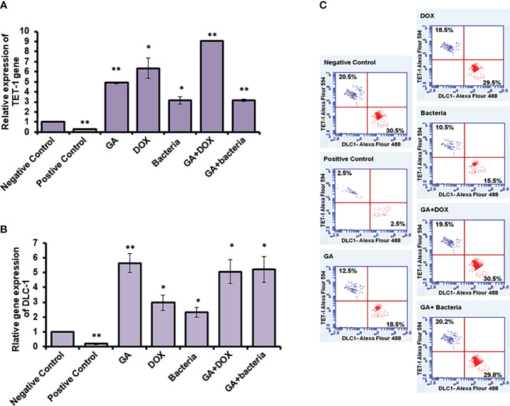
Method: In the current work, we investigated the effect of sole GA combined with different agents such as doxorubicin (DOX) or probiotic bacteria (Lactobacillus rhamanosus) against hepatocarcinogenesis induced by diethylnitrosamine to improve efficiency. The genomic DNA was isolated from rats' liver tissues to evaluate either methylation-sensitive or methylation-dependent resection enzymes. The methylation activity of the targeting genes DLC-1, TET-1, NF-kB, and STAT-3 was examined using specific primers and cleaved DNA products. Furthermore, flow cytometry was used to determine the protein expression profiles of DLC-1 and TET-1 in treated rats' liver tissue.

Results: Our results demonstrated the activity of GA to reduce the methylation activity in TET-1 and DLC-1 by 33.6% and 78%, respectively. As compared with the positive control. Furthermore, the association of GA with DOX avoided the methylation activity by 88% and 91% for TET-1 and DLC-1, respectively, as compared with the positive control. Similarly, the combined use of GA with probiotics suppressed the methylation activity in the TET-1 and DLC-1 genes by 75% and 81% for TET-1 and DLC-1, respectively. Also, GA and its combination with bacteria attenuated the adverse effect in hepatocarcinogenesis rats by altering potential methylomic genes such as NF-kb and STAT3 genes by 76% and 83%, respectively.

Conclusion: GA has an ameliorative effect against methylation inhibitors in hepatocellular carcinoma (HCC) by decreasing the methylation activity genes.

Keywords: 18β-Glycyrrhetinic acid (GA); DLC-1 and TET-1; NF-kB; STAT-3 methylation inhibitors; epigenetics; hepatocellular carcinoma (HCC); lactobacillus rhamanosus.


Conflict of interest statement

The authors declare that the research was conducted in the absence of any commercial or financial relationships that could be construed as a potential conflict of interest.

Figures

Figure 1
Figure 1
Chemical induction of HCC in albino rats treated with different effectors. (A) Body weight after treatment of HCC with different effectors. (B) Organ weight (lung, heart, kidney and liver) after treatment of HCC with different effectors.
Figure 2
Figure 2
Histological studies on evaluation of different compounds against HCC induced in albino rats A photomicrographic section of normal liver from the negative control group clearly shows normal liver morphology, whereas the positive control carcinogenic group demonstrates necrobiotic alterations and focal pleomorphic neoplastic cells, and rat liver treated with DOX illustrates mild swelling of hepatic carcinoma cells with hyperplasia of Kupffer cells. Poor <50% tumor necrosis, but with rat liver treated with GA showing necrobiotic changes of hepatic carcinoma cells with 51%-99% tumor necrosis, while also rat liver treated with GA and DOX showing small focal necrotic areas with apoptosis of a few hepatocytes, rat liver treated with bacteria showing vacuolar degeneration of hepatic carcinoma cells with hyperplasia of Kupffer cells and poor <50%percent tumor necrosis, and photomicrograph of rat liver section demonstrated necrobiotic changes of hepatic carcinoma cells. (A) Negative control animal. (B) positive control animals (rats with HCC). (C) The animal administrated received GA (100 mg/kg. orally) daily for 4 weeks. (D) Rats were injected with Doxorubicin (50 mg/kg) daily by i.p for 4 weeks. (E) The rats treated with probiotic bacteria (Lactobacillus rahmnosus) (2x10⁸ CFU/mL) were given it daily for 4 weeks. (F) The rats treated with Dox (50 mg/kg) with GA (100 mg/kg) daily for 4 weeks. (G) The rats were treated with probiotic bacteria (2x 10⁸ CFU/mL) with GA (100 mg/kg) daily for 4 weeks.
Figure 3
Figure 3
Measurement of the cofactor of methylation activity (DNMT1and MS) by qRT-PCR and flow cytometery. Positive control increased DNMT1 to 7-fold change while other treatment makes significant downregulation (A). Error bars indicate standard deviation of three independent experiments. The student’s two-tailed t-test was used for statistical analysis. P-value ≤ 0.05 was considered statistically significant. Positive control increased MS to 10-fold change while other treatment makes significant downregulation (B). Protein expression profile of DNMT1 and MS indicated by red and blue dotes using secondary antibodies Alex Flour-488 and 594, respectively (C). *statistically significant difference as compared with the controls (P, 0.05 for each). While, ** statistically significant difference as compared with the controls (P, 0.01 for each).
Figure 4
Figure 4
Methylation activity based on relative methylation in TET1 and DLC-1 in coding sequence and by restriction enzyme on the promoter region. (A) Fold change of methylated activity of TET-1 based on coding sequence. (B) Agarose gel electrophoresis of TET-1 gene segment that digested with CpG restriction enzyme HpaII indicates all derived samples in rats treated with GA, DOX, a combination of GA with bacteria and combination of GA with DOX in addition to controlling samples. (C) The methylation activity of DLC-1 is indicated by methylation fold change. The error bar indicates the stander deviation between two different replicates; statically, the student’s two-tails t-test test has been differentiating significantly. (D) Agarose gel electrophoresis of DLC-1 gene segment that digested with CpG restriction enzyme HpaII indicated all derived samples in rats treated with GA, DOX, a combination of GA with bacteria, and a combination of GA with DOX in addition to control samples. *statistically significant difference as compared with the controls (P, 0.05 for each). While, ** statistically significant difference as compared with the controls (P, 0.01 for each).
Figure 5
Figure 5
Relative methylation activity of NF-Kb and STAT3 based on RNA expression. (A, C) Relative methylation activity NF-kB and STAT-3 gene sequence indicated by fold change that significantly increased upon GA treatment and its combination with DOX or the bacteria. (B, D) relative expression of NF-kB and STAT-3 genes significantly reduced upon GA treatment and its indicated combination. Protein expression profile of NF-kB and STAT3 indicated by red and blue dotes using secondary antibodies Alex Flour-488 and 594, respectively (E). *statistically significant difference as compared with the controls (P, 0.05 for each). While, ** statistically significant difference as compared with the controls (P, 0.01 for each).
Figure 6
Figure 6
Relative expression of TET-1 and DLC-1 gene expression in GA and combination of GA with bacteria-treated rats. (A, B) Relative gene expression of TET-1 and DLC-1 indicated by fold change that was subjected to GA and combination of GA with Bacteria (lactobacillus rhamanosus) comparison with control. (C) Quantification of protein profile of TET-1 and DLC-1 in treated rats indicated by flow cytometry. *statistically significant difference as compared with the controls (P, 0.05 for each). While, ** statistically significant difference as compared with the controls (P, 0.01 for each).

References

    1. Hassan A, Elebeedy D, Matar ER, Fahmy Mohamed Elsayed A, Abd El Maksoud AI. Investigation of angiogenesis and wound healing potential mechanisms of Zinc Oxide Nanorods. Front Pharmacol (2021) 12:661217. doi: 10.3389/fphar.2021.661217 - DOI - PMC - PubMed
    1. Zhu RX, Seto WK, Lai CL, Yuen MF. Epidemiology of hepatocellular carcinoma in the Asia-Pacific region. Gut Liver (2016) 10:332–9. doi: 10.5009/gnl15257 - DOI - PMC - PubMed
    1. Bray F, Ferlay J, Soerjomataram I, Siegel RL, Torre LA, Jemal A. Global cancer statistics 2018: GLOBOCAN estimates of incidence and mortality worldwide for 36 cancers in 185 countries. CA Cancer J Clin (2018) 68:394–424. doi: 10.3322/caac.21492 - DOI - PubMed
    1. Kanda T, Goto T, Hirotsu Y, Moriyama M, Omata M. Molecular mechanisms driving progression of liver cirrhosis towards hepatocellular carcinoma in chronic hepatitis B and C infections: A review. Int J Mol Sci (2019) 20:1358. doi: 10.3390/ijms20061358 - DOI - PMC - PubMed
    1. Brown ZJ, Heinrich B, Greten TF. Mouse models of hepatocellular carcinoma: an overview and highlights for immunotherapy research. Nat Rev Gastroenterol Hepatol (2018) 15(9):536–54. doi: 10.1038/s41575-018-0033-6 - DOI - PubMed

Publication types