. 2024 Jan 23:14:1343503.
doi: 10.3389/fimmu.2023.1343503. eCollection 2023.

Bystander activation of microglia by Brucella abortus-infected astrocytes induces neuronal death via IL-6 trans-signaling

Affiliations

Bystander activation of microglia by Brucella abortus-infected astrocytes induces neuronal death via IL-6 trans-signaling

Julia Rodríguez et al. Front Immunol. .

Abstract

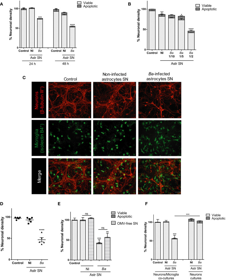
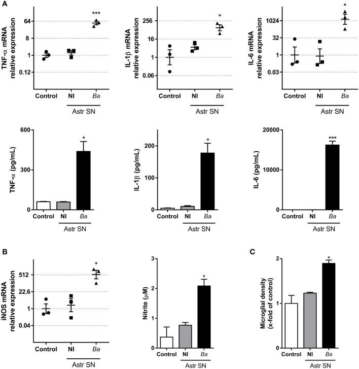
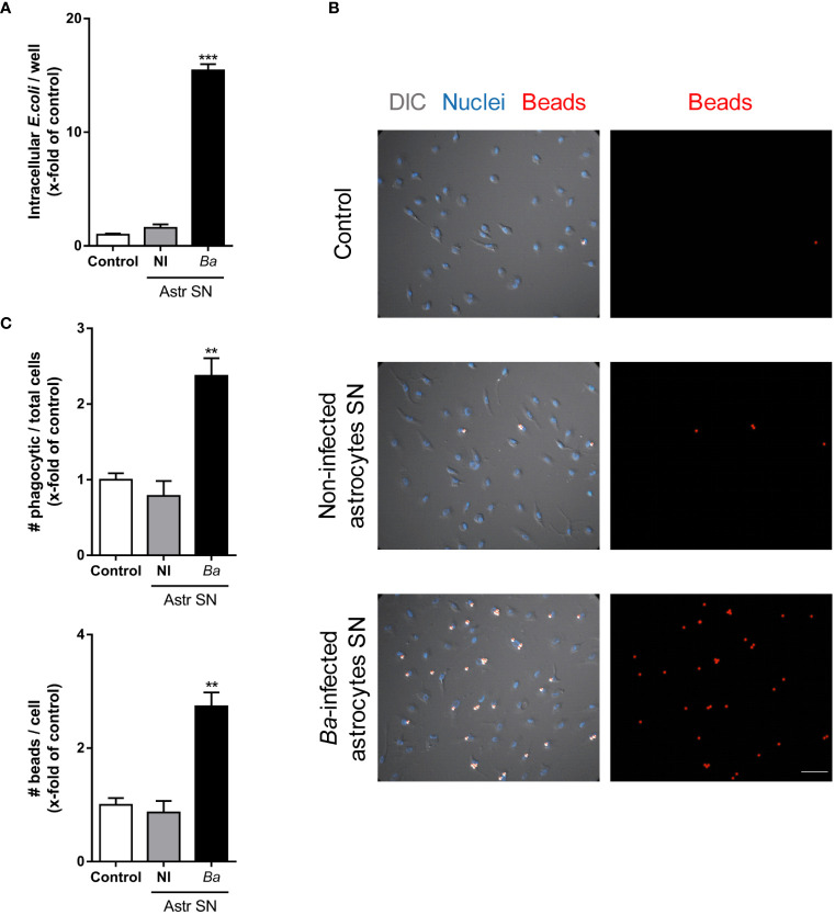
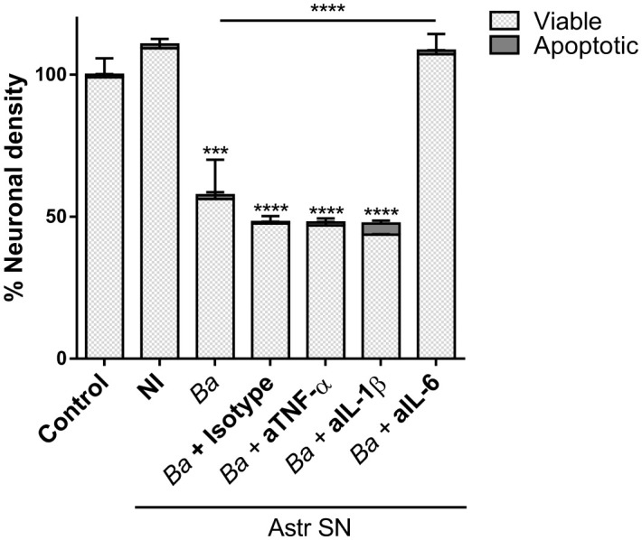
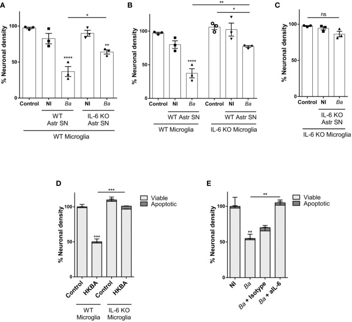
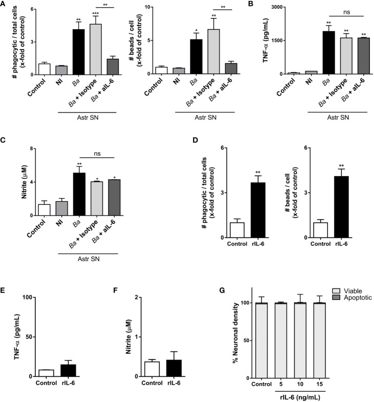
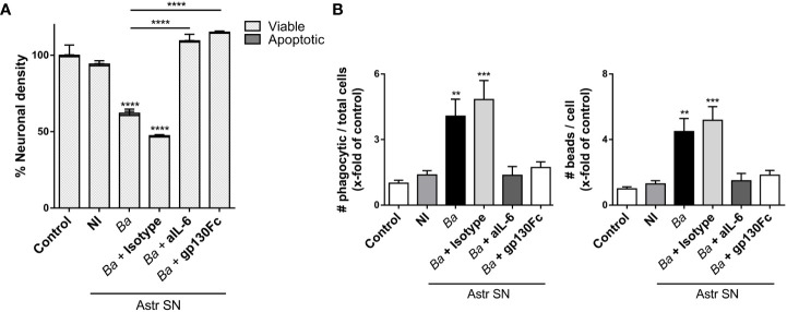
Inflammation plays a key role in the pathogenesis of neurobrucellosis where glial cell interactions are at the root of this pathological condition. In this study, we present evidence indicating that soluble factors secreted by Brucella abortus-infected astrocytes activate microglia to induce neuronal death. Culture supernatants (SN) from B. abortus-infected astrocytes induce the release of pro-inflammatory mediators and the increase of the microglial phagocytic capacity, which are two key features in the execution of live neurons by primary phagocytosis, a recently described mechanism whereby B. abortus-activated microglia kills neurons by phagocytosing them. IL-6 neutralization completely abrogates neuronal loss. IL-6 is solely involved in increasing the phagocytic capacity of activated microglia as induced by SN from B. abortus-infected astrocytes and does not participate in their inflammatory activation. Both autocrine microglia-derived and paracrine astrocyte-secreted IL-6 endow microglial cells with up-regulated phagocytic capacity that allows them to phagocytose neurons. Blocking of IL-6 signaling by soluble gp130 abrogates microglial phagocytosis and concomitant neuronal death, indicating that IL-6 activates microglia via trans-signaling. Altogether, these results demonstrate that soluble factors secreted by B. abortus-infected astrocytes activate microglia to induce, via IL-6 trans-signaling, the death of neurons. IL-6 signaling inhibition may thus be considered a strategy to control inflammation and CNS damage in neurobrucellosis.

Keywords: Brucella abortus; IL-6; astrocytes; microglia; neurobrucellosis; phagocytosis; trans-signaling.


Conflict of interest statement

The authors declare that the research was conducted in the absence of any commercial or financial relationships that could be construed as a potential conflict of interest.

Figures

Figure 1
Figure 1
SN from B. abortus-infected astrocytes induce neuronal death in co-cultures of neurons/microglia. Neurons/microglia co-cultures were stimulated with SN from non-infected (NI) or B. abortus-infected (Ba) astrocytes (Astr SN) diluted in complete medium (1/2) or left untreated (control) for 24 h or 48 h (A), or with different dilutions of SN from infected astrocytes for 48 h (B). Representative images from neurons/microglia co-cultures showing neurons labeled with anti-β-Tubulin III antibody (red) and microglia labeled with isolectin-B4 (green). Scale bar: 50 μm (C). The percentage (%) of neuronal density was evaluated in five independent experiments using neurons, microglia, and astrocytes SN from different animals (D). Astrocytes SN were ultracentrifuged (OMVs-free SN) or not and used to stimulate neurons/microglia co-cultures for 48 h (E). Cultures of neurons with microglia or neurons alone were treated with SN from non-infected or B. abortus-infected astrocytes for 48 h and neuronal density was evaluated (F). The density of neurons was evaluated by fluorescence microscopy. The percentage (%) of viable and apoptotic neurons was calculated vs. control condition. Data are shown as mean ± SEM from a representative experiment of three performed, except where indicated. *p < 0.05; **p < 0.005; ***p < 0.0005; ****p < 0.0001 vs. control condition, except where indicated. Non-significant (ns).
Figure 2
Figure 2
SN from B. abortus-infected astrocytes induce an inflammatory phenotype on microglia. Microglia were treated with SN from non-infected (NI) or B. abortus-infected (Ba) astrocytes (Astr SN) or left untreated (control) for 48 h. Gene expression of TNF-α, IL-1β, and IL-6 was analyzed by RT-qPCR in three independent experiments (plotted in a log2 scale) and cytokine secretion was measured by ELISA (A). Gene expression of iNOS was determined by RT-qPCR in three independent experiments and the level of NO was evaluated by Griess reaction (B). Proliferation was assessed by fluorescence microscopy (C). Data are shown as mean ± SEM from a representative experiment of three performed, except where indicated. *p < 0.05; ***p < 0.0005 vs. control condition.
Figure 3
Figure 3
SN from B. abortus-infected astrocytes increase the phagocytic activity of microglia. Microglia cultures were treated with SN from non-infected (NI) or B. abortus-infected (Ba) astrocytes (Astr SN), or left untreated (control). The phagocytic activity of microglia was evaluated by two different phagocytosis assays using E. coli (A) or negatively charged fluorescent 5 μm beads (B, C). Phagocytized bacteria were evaluated by intracellular CFU counting, and phagocytized beads were evaluated by fluorescence microscopy. Scale bar: 50 μm. Data are shown as mean ± SEM from a representative experiment of three performed. **p < 0.005; ***p < 0.0005 vs. control condition.
Figure 4
Figure 4
Microglia activated by B. abortus-infected astrocytes induce neuronal death by primary phagocytosis. Neurons/microglia co-cultures were stimulated with culture supernatants (Astr SN) from non-infected (NI) or B. abortus-infected (Ba) astrocytes for 48 h in the absence or the presence of recombinant Annexin V (rAnnexin V; 200 nM) (A), the cyclic (c) peptides cRAD or cRGD (100 μM) (B), or aminoguanidine (AG; 200 μM) (C). Untreated co-cultures were used as control conditions (control). The density of neurons was evaluated by fluorescence microscopy. The percentage (%) of viable and apoptotic neurons was calculated vs. control. Data are shown as mean ± SEM from a representative experiment of three performed. *p < 0.05; **p < 0.005; ***p < 0.0005 vs. control condition, except where indicated.
Figure 5
Figure 5
Neutralization of IL-6 prevents neuronal death induced by bystander-activated microglia. SN from non-infected (NI) or B. abortus-infected (Ba) astrocytes (Astr SN) were pre-incubated or not with anti-TNF-α (aTNF-α; 10 μg/mL), anti-IL-1β (aIL-1β; 10 μg/mL), anti-IL-6 (aIL-6; 5 μg/mL) monoclonal antibodies, or isotype control (10 μg/mL) and used to stimulate neurons/microglia co-cultures for 48 h. Untreated co-cultures were used as control conditions (control). The percentage (%) of viable and apoptotic neurons was calculated vs. control. Data are shown as mean ± SEM from a representative experiment of three performed. ***p < 0.0005; ****p < 0.0001 vs. control condition, except where indicated.
Figure 6
Figure 6
IL-6 secreted by both astrocytes and microglia contributes to neuronal death by primary phagocytosis. Neurons/microglia wild-type (WT) co-cultures were stimulated with SN from non-infected (NI) or B. abortus-infected (Ba) WT and IL-6 knock out (KO) astrocytes (Astr SN) for 48 h (A). Co-cultures of neurons and WT or IL-6 KO microglia were treated with SN from non-infected (NI) or (B) abortus-infected (Ba) WT astrocytes for 48 h (B). Co-cultures of WT neurons and IL-6 KO microglia were treated with SN from non-infected (NI) or (B) abortus-infected (Ba) IL-6 KO astrocytes for 48 h (C). Co-cultures of neurons and WT or IL-6 KO microglia were stimulated with heat-killed (B) abortus (HKBA; 1x10⁸ bacteria/mL) for 48 h (D). Neurons/microglia co-cultures were infected with B. abortus (MOI 100) in the presence of anti-IL-6 monoclonal antibody (aIL-6; 5 μg/mL) or its isotype control (5 μg/mL) for 48 h (E). The percentage (%) of neuronal density is shown as mean ± SEM from three independent experiments using neurons, microglia, and astrocytes SN from different animals (A-C). The percentage (%) of viable and apoptotic neurons was calculated vs. WT control (untreated) condition. Data are shown as mean ± SEM from a representative experiment of three performed (D, E). *p < 0.05; **p < 0.005; ***p < 0.0005; ****p < 0.0001 vs. WT control condition, except where indicated. Non-significant (ns).
Figure 7
Figure 7
IL-6 neutralization inhibits the phagocytic activity of microglia, but not its inflammatory activation. Microglia cultures were stimulated with SN from non-infected (NI) or B. abortus- infected (Ba) astrocytes (Astr SN) in the presence of anti-IL-6 monoclonal antibody (aIL-6; 5 μg/mL) or its isotype control (5 μg/mL) for 48 (h) Untreated co-culture was used as a control condition (control). Phagocytic activity was evaluated by a phagocytosis assay with fluorescent 5 μm beads that were visualized by fluorescence microscopy (A). Secretion of TNF-α (B) and NO (C) were also measured in cultured supernatants. Microglia cultures were treated with recombinant IL-6 (rIL-6; 15 ng/mL) for 48 h and phagocytic activity (D), secretion of TNF-α (E), and release of NO (F) were measured. Neurons/microglia co-cultures were treated with different concentrations of rIL-6 for 48 h, and neuronal density was evaluated. Untreated co-culture was used as a control condition (control). The percentage (%) of viable and apoptotic neurons was calculated vs. control (G). Data are shown as mean ± SEM from a representative experiment of three performed. *p < 0.05; **p < 0.005; ***p < 0.0005 vs. control condition, except where indicated. Non-significant (ns).
Figure 8
Figure 8
IL-6 activates microglia via trans-signaling. SN from non-infected (NI) or B. abortus-infected (Ba) astrocytes (Astr SN) were pre-incubated or not with recombinant gp130Fc (100 ng/mL), anti-IL-6 (aIL-6; 5 μg/mL) monoclonal antibody, or its isotype control (5 μg/mL) and used to stimulate neurons/microglia co-cultures (A) or microglia cultures (B) for 48 (h) Untreated cultures were used as control conditions (control). Percentage (%) of viable and apoptotic neurons was calculated vs. control (A). The phagocytic activity of microglia was evaluated (B). Data are shown as mean ± SEM from a representative experiment of three performed. **p < 0.005; ***p < 0.0005,****p < 0.0001 vs. control condition, except where indicated.

Similar articles

Cited by

References

    1. Pappas G, Akritidis N, Bosilkovski M, Tsianos E. Brucellosis. N Engl J Med (2005) 352(22):2325–36. doi: 10.1056/NEJMra050570 - DOI - PubMed
    1. Bouza E, Garcia de la Torre M, Parras F, Guerrero A, Rodriguez-Creixems M, Gobernado J. Brucellar meningitis. Rev Infect Dis (1987) 9(4):810–22. doi: 10.1093/clinids/9.4.810 - DOI - PubMed
    1. McLean DR, Russell N, Khan MY. Neurobrucellosis: clinical and therapeutic features. Clin Infect diseases: an Off Publ Infect Dis Soc America. (1992) 15(4):582–90. doi: 10.1093/clind/15.4.582 - DOI - PubMed
    1. Nalini A, Nagarathna S, Rajeshwari S, Rose D, Veena Kumari HB, Nagalingam M. Dementia, peripheral neuropathy, and chronic meningitis in Neurobrucellosis. Indian J Pathol Microbiol (2012) 55(1):128–30. doi: 10.4103/0377-4929.94896 - DOI - PubMed
    1. Shehata GA, Abdel-Baky L, Rashed H, Elamin H. Neuropsychiatric evaluation of patients with brucellosis. J neurovirology. (2010) 16(1):48–55. doi: 10.3109/13550280903586386 - DOI - PubMed

Publication types

LinkOut - more resources