

Editorial
doi: 10.7150/ijbs.92589.
eCollection 2024.
Digging out MDIG from the mess of H3K9me3, OTX2 and MYC signaling in human cancers
Affiliations
- PMID: 38322115
- PMCID: PMC10845298
- DOI: 10.7150/ijbs.92589
Item in Clipboard
Editorial
Digging out MDIG from the mess of H3K9me3, OTX2 and MYC signaling in human cancers
Int J Biol Sci.
.
No abstract available
Conflict of interest statement
Competing Interests: The authors have declared that no competing interest exists.
Figures
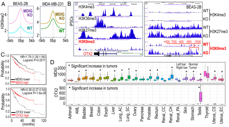
Antagonism of MDIG on histone H3 lysine 9 trimethylation (H3K9me3). A. ChIP-seq reveals that MDIG knockout enhances H3K9me3 enrichment on the genome of human bronchial epithelial cell line BEAS-2B cells (left) and human triple negative breast cancer cell line MDA-MB-231 cells (Right). B. OTX2 gene is enriched with peaks of active transcription markers (H3K4me3 and H3K36me3), but not repressive marker H3K9me3 in human induced pluripotent stem cells (iPSCs, Nips-B2). The bivalency of H3K27me3 and H3K4me3 indicates the poised status of OTX2 in iPSCs (left). Right panel: OTX2 gene is located in a transcriptionally inactive region on chromosome 14 featured with deminished H3K4me3 peaks in BEAS-2B cells. Knockout of MDIG (KO) enhanced the enrichment of H3K9me3 on OTX2 gene. Red lines and numbers indicate the positioning of H3K9me3 peaks in MDIG KO cells identified by ChIP-seq, which is similar to the findings by Du et al. in ChIP-PCR for the mouse OTX2 gene . C. Opposite effects of MDIG and OTX2 on HCC patient survival. High expression of MDIG predicts poorer survival (upper), whereas high expression of OTX2 predicts better survival of the patients with HCC (bottom). Data are derived from the Kaplan-Meier cancer patient survival and RNA-seq gene expression database by auto selection of the best cutoff values between the computed lower and upper quartiles. D. Significantly increased expression of MDIG in the indicated tumors relative to the case-matched normal tissues (upper), whereas except testis tumor, OTX2 is nearly undetectable in the indicated 21 types of tumors (bottom). Data are extrapolated from TNMplotter database using the relative expression values of MDIG and OTX2 mRNAs. (All data in this figure are from the authors' laboratory of this commentary).
References
-
- Lu Y, Chang Q, Zhang Y, Beezhold K, Rojanasakul Y, Zhao H. et al. Lung cancer-associated JmjC domain protein mdig suppresses formation of tri-methyl lysine 9 of histone H3. Cell Cycle. 2009;8:2101–9. - PubMed
Publication types
MeSH terms
Substances
Grants and funding
LinkOut - more resources
Full Text Sources
Medical
